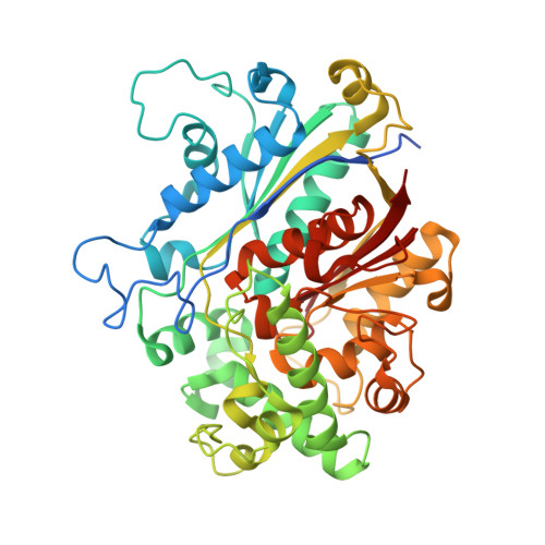
5LNQ

Crystal structure of SCP2 thiolase from Leishmania mexicana. Complex of the C123A mutant with acetoacetyl-CoA.


Experimental Data Snapshot

  • Method: X-RAY DIFFRACTION
  • Resolution: 1.98 Å
  • R-Value Free: 0.226 
  • R-Value Work: 0.189 
  • R-Value Observed: 0.191 

Starting Model: experimental
View more details

wwPDB Validation   3D Report Full Report


Ligand Structure Quality Assessment 


This is version 1.2 of the entry. See complete history


Literature

Crystallographic substrate binding studies of Leishmania mexicana SCP2-thiolase (type-2): unique features of oxyanion hole-1.

Harijan, R.K.Kiema, T.R.Syed, S.M.Qadir, I.Mazet, M.Bringaud, F.Michels, P.A.M.Wierenga, R.K.

(2017) Protein Eng Des Sel 30: 225-233

  • DOI: https://doi.org/10.1093/protein/gzw080
  • Primary Citation of Related Structures:  
    5LNQ, 5LOT

  • PubMed Abstract: 

    Structures of the C123A variant of the dimeric Leishmania mexicana SCP2-thiolase (type-2) (Lm-thiolase), complexed with acetyl-CoA and acetoacetyl-CoA, respectively, are reported. The catalytic site of thiolase contains two oxyanion holes, OAH1 and OAH2, which are important for catalysis. The two structures reveal for the first time the hydrogen bond interactions of the CoA-thioester oxygen atom of the substrate with the hydrogen bond donors of OAH1 of a CHH-thiolase. The amino acid sequence fingerprints ( xS, EAF, G P) of three catalytic loops identify the active site geometry of the well-studied CNH-thiolases, whereas SCP2-thiolases (type-1, type-2) are classified as CHH-thiolases, having as corresponding fingerprints xS, DCF and G P. In all thiolases, OAH2 is formed by the main chain NH groups of two catalytic loops. In the well-studied CNH-thiolases, OAH1 is formed by a water (of the Wat-Asn(NEAF) dyad) and NE2 (of the GHP-histidine). In the two described liganded Lm-thiolase structures, it is seen that in this CHH-thiolase, OAH1 is formed by NE2 of His338 (HDCF) and His388 (GHP). Analysis of the OAH1 hydrogen bond networks suggests that the GHP-histidine is doubly protonated and positively charged in these complexes, whereas the HDCF histidine is neutral and singly protonated.


  • Organizational Affiliation

    Faculty of Biochemistry and Molecular Medicine, Biocenter Oulu, University of Oulu, FIN-90014, Finland.


Macromolecules
Find similar proteins by:  (by identity cutoff)  |  3D Structure
Entity ID: 1
MoleculeChains Sequence LengthOrganismDetailsImage
3-ketoacyl-CoA thiolase-like protein
A, B, C, D
457Leishmania mexicanaMutation(s): 1 
Gene Names: LMXM_23_0690
EC: 2.3.1.16 (PDB Primary Data), 2.3.1.176 (UniProt)
UniProt
Find proteins for E9AW84 (Leishmania mexicana (strain MHOM/GT/2001/U1103))
Explore E9AW84 
Go to UniProtKB:  E9AW84
Entity Groups  
Sequence Clusters30% Identity50% Identity70% Identity90% Identity95% Identity100% Identity
UniProt GroupE9AW84
Sequence Annotations
Expand
  • Reference Sequence
Experimental Data & Validation

Experimental Data

  • Method: X-RAY DIFFRACTION
  • Resolution: 1.98 Å
  • R-Value Free: 0.226 
  • R-Value Work: 0.189 
  • R-Value Observed: 0.191 
  • Space Group: P 1 21 1
Unit Cell:
Length ( Å )Angle ( ˚ )
a = 74.141α = 90
b = 89.804β = 104.76
c = 123.716γ = 90
Software Package:
Software NamePurpose
REFMACrefinement
XDSdata reduction
Aimlessdata scaling
PHASERphasing

Structure Validation

View Full Validation Report


Ligand Structure Quality Assessment 


Entry History & Funding Information

Deposition Data


Funding OrganizationLocationGrant Number
Academy of FinlandFinland131795
BioStruct-XFinland283570
European Commissions Seventh Framework ProgrammeFinlandFP7/2007-2013

Revision History  (Full details and data files)

  • Version 1.0: 2017-01-18
    Type: Initial release
  • Version 1.1: 2017-05-03
    Changes: Database references
  • Version 1.2: 2024-01-10
    Changes: Data collection, Database references, Refinement description