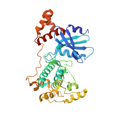
5MAF

Crystal structure of MELK in complex with an inhibitor


Experimental Data Snapshot

  • Method: X-RAY DIFFRACTION
  • Resolution: 2.80 Å
  • R-Value Free: 0.259 
  • R-Value Work: 0.210 
  • R-Value Observed: 0.212 

Starting Model: other
View more details

wwPDB Validation   3D Report Full Report


Ligand Structure Quality Assessment 


This is version 1.3 of the entry. See complete history


Literature

The target landscape of clinical kinase drugs.

Klaeger, S.Heinzlmeir, S.Wilhelm, M.Polzer, H.Vick, B.Koenig, P.A.Reinecke, M.Ruprecht, B.Petzoldt, S.Meng, C.Zecha, J.Reiter, K.Qiao, H.Helm, D.Koch, H.Schoof, M.Canevari, G.Casale, E.Depaolini, S.R.Feuchtinger, A.Wu, Z.Schmidt, T.Rueckert, L.Becker, W.Huenges, J.Garz, A.K.Gohlke, B.O.Zolg, D.P.Kayser, G.Vooder, T.Preissner, R.Hahne, H.Tonisson, N.Kramer, K.Gotze, K.Bassermann, F.Schlegl, J.Ehrlich, H.C.Aiche, S.Walch, A.Greif, P.A.Schneider, S.Felder, E.R.Ruland, J.Medard, G.Jeremias, I.Spiekermann, K.Kuster, B.

(2017) Science 358

  • DOI: https://doi.org/10.1126/science.aan4368
  • Primary Citation of Related Structures:  
    5LBW, 5LBY, 5LBZ, 5M5A, 5MAF, 5MAG, 5MAH, 5MAI

  • PubMed Abstract: 

    Kinase inhibitors are important cancer therapeutics. Polypharmacology is commonly observed, requiring thorough target deconvolution to understand drug mechanism of action. Using chemical proteomics, we analyzed the target spectrum of 243 clinically evaluated kinase drugs. The data revealed previously unknown targets for established drugs, offered a perspective on the "druggable" kinome, highlighted (non)kinase off-targets, and suggested potential therapeutic applications. Integration of phosphoproteomic data refined drug-affected pathways, identified response markers, and strengthened rationale for combination treatments. We exemplify translational value by discovering SIK2 (salt-inducible kinase 2) inhibitors that modulate cytokine production in primary cells, by identifying drugs against the lung cancer survival marker MELK (maternal embryonic leucine zipper kinase), and by repurposing cabozantinib to treat FLT3-ITD-positive acute myeloid leukemia. This resource, available via the ProteomicsDB database, should facilitate basic, clinical, and drug discovery research and aid clinical decision-making.


  • Organizational Affiliation

    Chair of Proteomics and Bioanalytics, Technical University of Munich (TUM), Freising, Germany.


Macromolecules
Find similar proteins by:  (by identity cutoff)  |  3D Structure
Entity ID: 1
MoleculeChains Sequence LengthOrganismDetailsImage
Maternal embryonic leucine zipper kinase347Homo sapiensMutation(s): 0 
Gene Names: MELKKIAA0175
EC: 2.7.11.1 (PDB Primary Data), 2.7.10.2 (PDB Primary Data)
UniProt & NIH Common Fund Data Resources
Find proteins for Q14680 (Homo sapiens)
Explore Q14680 
Go to UniProtKB:  Q14680
PHAROS:  Q14680
GTEx:  ENSG00000165304 
Entity Groups  
Sequence Clusters30% Identity50% Identity70% Identity90% Identity95% Identity100% Identity
UniProt GroupQ14680
Sequence Annotations
Expand
  • Reference Sequence
Small Molecules
Ligands 3 Unique
IDChains Name / Formula / InChI Key2D Diagram3D Interactions
XIN
Query on XIN

Download Ideal Coordinates CCD File 
D [auth A]methyl (3Z)-3-{[(4-{methyl[(4-methylpiperazin-1-yl)acetyl]amino}phenyl)amino](phenyl)methylidene}-2-oxo-2,3-dihydro-1H-indole-6-carboxylate
C31 H33 N5 O4
XZXHXSATPCNXJR-ZIADKAODSA-N
DMS
Query on DMS

Download Ideal Coordinates CCD File 
B [auth A]DIMETHYL SULFOXIDE
C2 H6 O S
IAZDPXIOMUYVGZ-UHFFFAOYSA-N
CL
Query on CL

Download Ideal Coordinates CCD File 
C [auth A]CHLORIDE ION
Cl
VEXZGXHMUGYJMC-UHFFFAOYSA-M
Experimental Data & Validation

Experimental Data

  • Method: X-RAY DIFFRACTION
  • Resolution: 2.80 Å
  • R-Value Free: 0.259 
  • R-Value Work: 0.210 
  • R-Value Observed: 0.212 
  • Space Group: P 21 21 21
Unit Cell:
Length ( Å )Angle ( ˚ )
a = 59.913α = 90
b = 63.713β = 90
c = 91.292γ = 90
Software Package:
Software NamePurpose
REFMACrefinement
XDSdata reduction
SCALAdata scaling
PHASERphasing

Structure Validation

View Full Validation Report


Ligand Structure Quality Assessment 


Entry History 

Deposition Data

Revision History  (Full details and data files)

  • Version 1.0: 2017-12-06
    Type: Initial release
  • Version 1.1: 2017-12-13
    Changes: Database references
  • Version 1.2: 2024-05-01
    Changes: Data collection, Database references, Refinement description
  • Version 1.3: 2024-11-06
    Changes: Structure summary