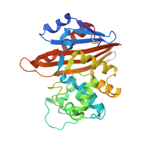
5MOZ

OXA-10 Avibactam complex with bound Iodide


Experimental Data Snapshot

  • Method: X-RAY DIFFRACTION
  • Resolution: 1.34 Å
  • R-Value Free: 0.185 
  • R-Value Work: 0.168 

Starting Model: experimental
View more details

wwPDB Validation   3D Report Full Report


Ligand Structure Quality Assessment 


This is version 1.3 of the entry. See complete history


Literature

(13)C-Carbamylation as a mechanistic probe for the inhibition of class D beta-lactamases by avibactam and halide ions.

Lohans, C.T.Wang, D.Y.Jorgensen, C.Cahill, S.T.Clifton, I.J.McDonough, M.A.Oswin, H.P.Spencer, J.Domene, C.Claridge, T.D.W.Brem, J.Schofield, C.J.

(2017) Org Biomol Chem 15: 6024-6032

  • DOI: https://doi.org/10.1039/c7ob01514c
  • Primary Citation of Related Structures:  
    5MMY, 5MNU, 5MOX, 5MOZ

  • PubMed Abstract: 

    The class D (OXA) serine β-lactamases are a major cause of resistance to β-lactam antibiotics. The class D enzymes are unique amongst β-lactamases because they have a carbamylated lysine that acts as a general acid/base in catalysis. Previous crystallographic studies led to the proposal that β-lactamase inhibitor avibactam targets OXA enzymes in part by promoting decarbamylation. Similarly, halide ions are proposed to inhibit OXA enzymes via decarbamylation. NMR analyses, in which the carbamylated lysines of OXA-10, -23 and -48 were 13 C-labelled, indicate that reaction with avibactam does not ablate lysine carbamylation in solution. While halide ions did not decarbamylate the 13 C-labelled OXA enzymes in the absence of substrate or inhibitor, avibactam-treated OXA enzymes were susceptible to decarbamylation mediated by halide ions, suggesting halide ions may inhibit OXA enzymes by promoting decarbamylation of acyl-enzyme complex. Crystal structures of the OXA-10 avibactam complex were obtained with bromide, iodide, and sodium ions bound between Trp-154 and Lys-70. Structures were also obtained wherein bromide and iodide ions occupy the position expected for the 'hydrolytic water' molecule. In contrast with some solution studies, Lys-70 was decarbamylated in these structures. These results reveal clear differences between crystallographic and solution studies on the interaction of class D β-lactamases with avibactam and halides, and demonstrate the utility of 13 C-NMR for studying lysine carbamylation in solution.


  • Organizational Affiliation

    Department of Chemistry, University of Oxford, Oxford, OX1 3TA, UK. [email protected] [email protected].


Macromolecules
Find similar proteins by:  (by identity cutoff)  |  3D Structure
Entity ID: 1
MoleculeChains Sequence LengthOrganismDetailsImage
Beta-lactamase OXA-10
A, B
247Pseudomonas aeruginosaMutation(s): 0 
Gene Names: blaoxa10pse2
EC: 3.5.2.6
UniProt
Find proteins for P14489 (Pseudomonas aeruginosa)
Explore P14489 
Go to UniProtKB:  P14489
Entity Groups  
Sequence Clusters30% Identity50% Identity70% Identity90% Identity95% Identity100% Identity
UniProt GroupP14489
Sequence Annotations
Expand
  • Reference Sequence
Small Molecules
Ligands 5 Unique
IDChains Name / Formula / InChI Key2D Diagram3D Interactions
NXL
Query on NXL

Download Ideal Coordinates CCD File 
C [auth A],
Y [auth B]
(2S,5R)-1-formyl-5-[(sulfooxy)amino]piperidine-2-carboxamide
C7 H13 N3 O6 S
WJDGWXPPFHLLNL-RITPCOANSA-N
IOD
Query on IOD

Download Ideal Coordinates CCD File 
N [auth A]
O [auth A]
P [auth A]
Q [auth A]
U [auth B]
N [auth A],
O [auth A],
P [auth A],
Q [auth A],
U [auth B],
V [auth B],
W [auth B],
X [auth B]
IODIDE ION
I
XMBWDFGMSWQBCA-UHFFFAOYSA-M
GOL
Query on GOL

Download Ideal Coordinates CCD File 
D [auth A]
E [auth A]
F [auth A]
G [auth A]
H [auth A]
D [auth A],
E [auth A],
F [auth A],
G [auth A],
H [auth A],
R [auth B]
GLYCEROL
C3 H8 O3
PEDCQBHIVMGVHV-UHFFFAOYSA-N
CL
Query on CL

Download Ideal Coordinates CCD File 
T [auth B]CHLORIDE ION
Cl
VEXZGXHMUGYJMC-UHFFFAOYSA-M
NA
Query on NA

Download Ideal Coordinates CCD File 
I [auth A]
J [auth A]
K [auth A]
L [auth A]
M [auth A]
I [auth A],
J [auth A],
K [auth A],
L [auth A],
M [auth A],
S [auth B]
SODIUM ION
Na
FKNQFGJONOIPTF-UHFFFAOYSA-N
Experimental Data & Validation

Experimental Data

  • Method: X-RAY DIFFRACTION
  • Resolution: 1.34 Å
  • R-Value Free: 0.185 
  • R-Value Work: 0.168 
  • Space Group: P 21 21 21
Unit Cell:
Length ( Å )Angle ( ˚ )
a = 48.841α = 90
b = 101.658β = 90
c = 126.727γ = 90
Software Package:
Software NamePurpose
PHENIXrefinement
PDB_EXTRACTdata extraction
PHASERphasing
XDSdata reduction
PHASERphasing

Structure Validation

View Full Validation Report


Ligand Structure Quality Assessment 


Entry History 

Deposition Data

  • Released Date: 2017-07-12 
  • Deposition Author(s): Brem, J.

Revision History  (Full details and data files)

  • Version 1.0: 2017-07-12
    Type: Initial release
  • Version 1.1: 2017-07-26
    Changes: Database references
  • Version 1.2: 2024-01-17
    Changes: Data collection, Database references, Derived calculations, Refinement description, Structure summary
  • Version 1.3: 2024-10-09
    Changes: Structure summary