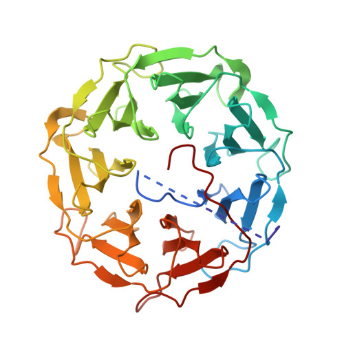
5MXH

Photorhabdus asymbiotica lectin (PHL) in complex with D-galactose


Experimental Data Snapshot

  • Method: X-RAY DIFFRACTION
  • Resolution: 1.95 Å
  • R-Value Free: 0.217 
  • R-Value Work: 0.179 
  • R-Value Observed: 0.181 

Starting Model: experimental
View more details

wwPDB Validation   3D Report Full Report


Ligand Structure Quality Assessment 


This is version 1.4 of the entry. See complete history


Literature

Characterization of novel bangle lectin from Photorhabdus asymbiotica with dual sugar-binding specificity and its effect on host immunity.

Jancarikova, G.Houser, J.Dobes, P.Demo, G.Hyrsl, P.Wimmerova, M.

(2017) PLoS Pathog 13: e1006564-e1006564

  • DOI: https://doi.org/10.1371/journal.ppat.1006564
  • Primary Citation of Related Structures:  
    5MXE, 5MXF, 5MXG, 5MXH

  • PubMed Abstract: 

    Photorhabdus asymbiotica is one of the three recognized species of the Photorhabdus genus, which consists of gram-negative bioluminescent bacteria belonging to the family Morganellaceae. These bacteria live in a symbiotic relationship with nematodes from the genus Heterorhabditis, together forming a complex that is highly pathogenic for insects. Unlike other Photorhabdus species, which are strictly entomopathogenic, P. asymbiotica is unique in its ability to act as an emerging human pathogen. Analysis of the P. asymbiotica genome identified a novel fucose-binding lectin designated PHL with a strong sequence similarity to the recently described P. luminescens lectin PLL. Recombinant PHL exhibited high affinity for fucosylated carbohydrates and the unusual disaccharide 3,6-O-Me2-Glcβ1-4(2,3-O-Me2)Rhaα-O-(p-C6H4)-OCH2CH2NH2 from Mycobacterium leprae. Based on its crystal structure, PHL forms a seven-bladed β-propeller assembling into a homo-dimer with an inter-subunit disulfide bridge. Investigating complexes with different ligands revealed the existence of two sets of binding sites per monomer-the first type prefers l-fucose and its derivatives, whereas the second type can bind d-galactose. Based on the sequence analysis, PHL could contain up to twelve binding sites per monomer. PHL was shown to interact with all types of red blood cells and insect haemocytes. Interestingly, PHL inhibited the production of reactive oxygen species induced by zymosan A in human blood and antimicrobial activity both in human blood, serum and insect haemolymph. Concurrently, PHL increased the constitutive level of oxidants in the blood and induced melanisation in haemolymph. Our results suggest that PHL might play a crucial role in the interaction of P. asymbiotica with both human and insect hosts.


  • Organizational Affiliation

    Central European Institute of Technology (CEITEC), Masaryk University, Brno, Czech Republic.


Macromolecules
Find similar proteins by:  (by identity cutoff)  |  3D Structure
Entity ID: 1
MoleculeChains Sequence LengthOrganismDetailsImage
Photorhabdus asymbiotica lectin PHL369Photorhabdus asymbiotica subsp. asymbiotica ATCC 43949Mutation(s): 0 
Gene Names: PAU_00698
UniProt
Find proteins for C7BLE4 (Photorhabdus asymbiotica subsp. asymbiotica (strain ATCC 43949 / 3105-77))
Explore C7BLE4 
Go to UniProtKB:  C7BLE4
Entity Groups  
Sequence Clusters30% Identity50% Identity70% Identity90% Identity95% Identity100% Identity
UniProt GroupC7BLE4
Sequence Annotations
Expand
  • Reference Sequence
Small Molecules
Ligands 3 Unique
IDChains Name / Formula / InChI Key2D Diagram3D Interactions
GAL
Query on GAL

Download Ideal Coordinates CCD File 
B [auth A],
C [auth A],
D [auth A]
beta-D-galactopyranose
C6 H12 O6
WQZGKKKJIJFFOK-FPRJBGLDSA-N
CL
Query on CL

Download Ideal Coordinates CCD File 
F [auth A],
G [auth A],
H [auth A],
I [auth A]
CHLORIDE ION
Cl
VEXZGXHMUGYJMC-UHFFFAOYSA-M
MG
Query on MG

Download Ideal Coordinates CCD File 
E [auth A]MAGNESIUM ION
Mg
JLVVSXFLKOJNIY-UHFFFAOYSA-N
Experimental Data & Validation

Experimental Data

  • Method: X-RAY DIFFRACTION
  • Resolution: 1.95 Å
  • R-Value Free: 0.217 
  • R-Value Work: 0.179 
  • R-Value Observed: 0.181 
  • Space Group: P 31 2 1
Unit Cell:
Length ( Å )Angle ( ˚ )
a = 81.323α = 90
b = 81.323β = 90
c = 114.181γ = 120
Software Package:
Software NamePurpose
REFMACrefinement
XDSdata reduction
SCALAdata scaling
PHASERphasing

Structure Validation

View Full Validation Report


Ligand Structure Quality Assessment 


Entry History 

Deposition Data

Revision History  (Full details and data files)

  • Version 1.0: 2017-08-09
    Type: Initial release
  • Version 1.1: 2017-08-23
    Changes: Database references
  • Version 1.2: 2020-07-29
    Type: Remediation
    Reason: Carbohydrate remediation
    Changes: Data collection, Derived calculations, Structure summary
  • Version 1.3: 2024-01-17
    Changes: Data collection, Database references, Refinement description, Structure summary
  • Version 1.4: 2024-11-13
    Changes: Structure summary