5N6Y

Azotobacter vinelandii vanadium nitrogenase


Experimental Data Snapshot

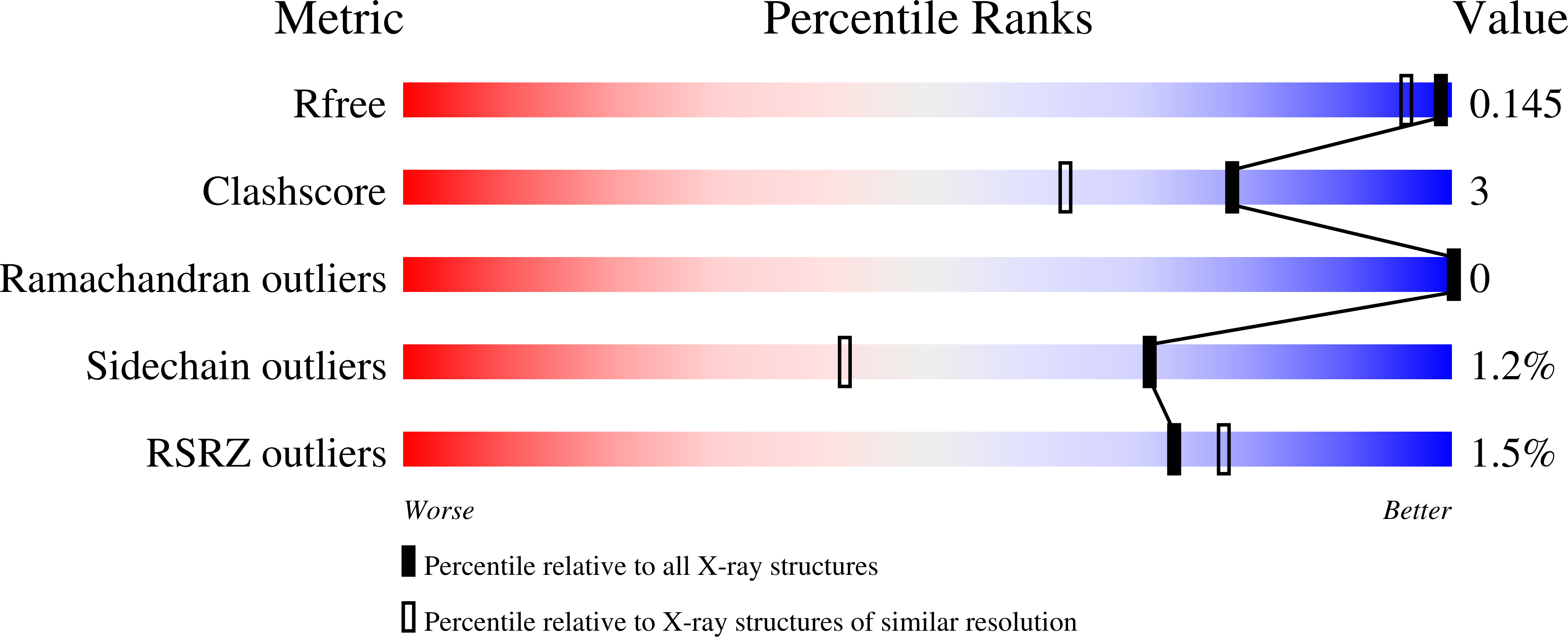
  • Method: X-RAY DIFFRACTION
  • Resolution: 1.35 Å
  • R-Value Free: 0.146 
  • R-Value Work: 0.109 
  • R-Value Observed: 0.111 

wwPDB Validation   3D Report Full Report


Ligand Structure Quality Assessment 


This is version 2.1 of the entry. See complete history


Literature

The structure of vanadium nitrogenase reveals an unusual bridging ligand.

Sippel, D.Einsle, O.

(2017) Nat Chem Biol 13: 956-960

  • DOI: https://doi.org/10.1038/nchembio.2428
  • Primary Citation of Related Structures:  
    5N6Y

  • PubMed Abstract: 

    Nitrogenases catalyze the reduction of dinitrogen (N 2 ) gas to ammonium at a complex heterometallic cofactor. This most commonly occurs at the FeMo cofactor (FeMoco), a [Mo-7Fe-9S-C] cluster whose exact reactivity and substrate-binding mode remain unknown. Alternative nitrogenases replace molybdenum with either vanadium or iron and differ in reactivity, most prominently in the ability of vanadium nitrogenase to reduce CO to hydrocarbons. Here we report the 1.35-Å structure of vanadium nitrogenase from Azotobacter vinelandii. The 240-kDa protein contains an additional α-helical subunit that is not present in molybdenum nitrogenase. The FeV cofactor (FeVco) is a [V-7Fe-8S-C] cluster with a homocitrate ligand to vanadium. Unexpectedly, it lacks one sulfide ion compared to FeMoco, which is replaced by a bridging ligand, likely a μ-1,3-carbonate. The anion fits into a pocket within the protein that is obstructed in molybdenum nitrogenase, and its different chemical character helps to rationalize the altered chemical properties of this unique N 2 - and CO-fixing enzyme.


  • Organizational Affiliation

    Lehrstuhl Biochemie, Institut für Biochemie, Albert-Ludwigs-Universität Freiburg, Freiburg Research Institute for Advanced Studies (FRIAS), and BIOSS Centre for Biological Signalling Studies, Freiburg, Germany.


Macromolecules
Find similar proteins by:  (by identity cutoff)  |  3D Structure
Entity ID: 1
MoleculeChains Sequence LengthOrganismDetailsImage
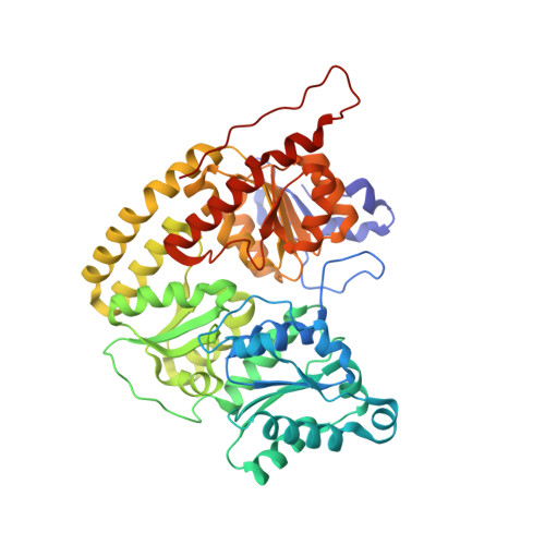
Nitrogenase vanadium-iron protein alpha chain
A, D
474Azotobacter vinelandiiMutation(s): 0 
Gene Names: vnfD
EC: 1.18.6.1
UniProt
Find proteins for P16855 (Azotobacter vinelandii)
Explore P16855 
Go to UniProtKB:  P16855
Entity Groups  
Sequence Clusters30% Identity50% Identity70% Identity90% Identity95% Identity100% Identity
UniProt GroupP16855
Sequence Annotations
Expand
  • Reference Sequence
Find similar proteins by:  (by identity cutoff)  |  3D Structure
Entity ID: 2
MoleculeChains Sequence LengthOrganismDetailsImage
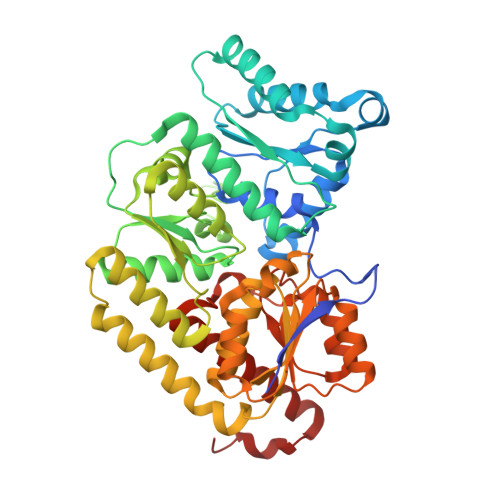
Nitrogenase vanadium-iron protein beta chainB [auth E],
E [auth B]
475Azotobacter vinelandiiMutation(s): 0 
Gene Names: vnfK
EC: 1.18.6.1
UniProt
Find proteins for P16856 (Azotobacter vinelandii)
Explore P16856 
Go to UniProtKB:  P16856
Entity Groups  
Sequence Clusters30% Identity50% Identity70% Identity90% Identity95% Identity100% Identity
UniProt GroupP16856
Sequence Annotations
Expand
  • Reference Sequence
Find similar proteins by:  (by identity cutoff)  |  3D Structure
Entity ID: 3
MoleculeChains Sequence LengthOrganismDetailsImage
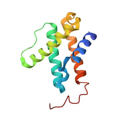
Nitrogenase vanadium-iron protein delta chain
C, F
113Azotobacter vinelandiiMutation(s): 0 
Gene Names: vnfG
EC: 1.18.6.1
UniProt
Find proteins for P16857 (Azotobacter vinelandii)
Explore P16857 
Go to UniProtKB:  P16857
Entity Groups  
Sequence Clusters30% Identity50% Identity70% Identity90% Identity95% Identity100% Identity
UniProt GroupP16857
Sequence Annotations
Expand
  • Reference Sequence
Small Molecules
Ligands 5 Unique
IDChains Name / Formula / InChI Key2D Diagram3D Interactions
8P8
Query on 8P8

Download Ideal Coordinates CCD File 
H [auth A],
P [auth D]
C Fe7 S8 V
C Fe7 S8 V
DYVZWQKCGNUCRR-UHFFFAOYSA-N
CLF
Query on CLF

Download Ideal Coordinates CCD File 
J [auth A],
K [auth E]
FE(8)-S(7) CLUSTER
Fe8 S7
JKVMXLBGZBULKV-UHFFFAOYSA-N
HCA
Query on HCA

Download Ideal Coordinates CCD File 
G [auth A],
O [auth D]
3-HYDROXY-3-CARBOXY-ADIPIC ACID
C7 H10 O7
XKJVEVRQMLKSMO-SSDOTTSWSA-N
CO3
Query on CO3

Download Ideal Coordinates CCD File 
I [auth A],
Q [auth D]
CARBONATE ION
C O3
BVKZGUZCCUSVTD-UHFFFAOYSA-L
MG
Query on MG

Download Ideal Coordinates CCD File 
L [auth E],
M [auth E],
N [auth C],
R [auth F]
MAGNESIUM ION
Mg
JLVVSXFLKOJNIY-UHFFFAOYSA-N
Experimental Data & Validation

Experimental Data

  • Method: X-RAY DIFFRACTION
  • Resolution: 1.35 Å
  • R-Value Free: 0.146 
  • R-Value Work: 0.109 
  • R-Value Observed: 0.111 
  • Space Group: P 1
Unit Cell:
Length ( Å )Angle ( ˚ )
a = 75.25α = 84.06
b = 79.79β = 72.62
c = 106.97γ = 75.15
Software Package:
Software NamePurpose
REFMACrefinement
XDSdata reduction
SCALAdata scaling
PHENIXphasing

Structure Validation

View Full Validation Report


Ligand Structure Quality Assessment 


Entry History & Funding Information

Deposition Data


Funding OrganizationLocationGrant Number
European Research CouncilGermany310656
German Research FoundationGermanyRTG 1976
German Research FoundationGermanyEI-520/10

Revision History  (Full details and data files)

  • Version 1.0: 2017-07-26
    Type: Initial release
  • Version 1.1: 2017-08-30
    Changes: Database references
  • Version 2.0: 2017-09-20
    Changes: Advisory, Atomic model, Database references, Derived calculations, Polymer sequence
  • Version 2.1: 2024-05-08
    Changes: Data collection, Database references, Derived calculations