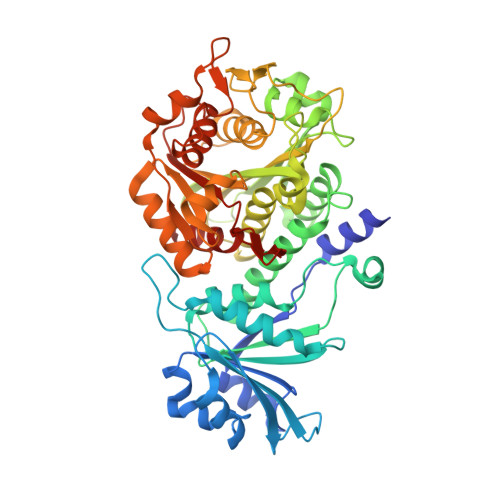
Structural Characterization of Acidic M17 Leucine Aminopeptidases from the TriTryps and Evaluation of Their Role in Nutrient Starvation inTrypanosoma brucei.
Timm, J., Valente, M., Garcia-Caballero, D., Wilson, K.S., Gonzalez-Pacanowska, D.(2017) mSphere 2
- PubMed: 28815215
- DOI: https://doi.org/10.1128/mSphere.00226-17
- Primary Citation of Related Structures:
5NSK, 5NSM, 5NSQ, 5NTD, 5NTF, 5NTG, 5NTH - PubMed Abstract:
Leucine aminopeptidase (LAP) is found in all kingdoms of life and catalyzes the metal-dependent hydrolysis of the N-terminal amino acid residue of peptide or amino acyl substrates. LAPs have been shown to participate in the N-terminal processing of certain proteins in mammalian cells and in homologous recombination and transcription regulation in bacteria, while in parasites, they are involved in host cell invasion and provision of essential amino acids for growth. The enzyme is essential for survival in Plasmodium falciparum , where its drug target potential has been suggested. We report here the X-ray structures of three kinetoplastid acidic LAPs (LAP-As from Trypanosoma brucei , Trypanosoma cruzi , and Leishmania major ) which were solved in the metal-free and unliganded forms, as well as in a number of ligand complexes, providing insight into ligand binding, metal ion requirements, and oligomeric state. In addition, we analyzed mutant cells defective in LAP-A in Trypanosoma brucei , strongly suggesting that the enzyme is not required for the growth of this parasite either in vitro or in vivo . In procyclic cells, LAP-A was equally distributed throughout the cytoplasm, yet upon starvation, it relocalizes in particles that concentrate in the perinuclear region. Overexpression of the enzyme conferred a growth advantage when parasites were grown in leucine-deficient medium. Overall, the results suggest that in T. brucei , LAP-A may participate in protein degradation associated with nutrient depletion. IMPORTANCE Leucine aminopeptidases (LAPs) catalyze the hydrolysis of the N-terminal amino acid of peptides and are considered potential drug targets. They are involved in multiple functions ranging from host cell invasion and provision of essential amino acids to site-specific homologous recombination and transcription regulation. In kinetoplastid parasites, there are at least three distinct LAPs. The availability of the crystal structures provides important information for drug design. Here we report the structure of the acidic LAPs from three kinetoplastids in complex with different inhibitors and explore their role in Trypanosoma brucei survival under various nutrient conditions. Importantly, the acidic LAP is dispensable for growth both in vitro and in vivo , an observation that questions its use as a specific drug target. While LAP-A is not essential, leucine depletion and subcellular localization studies performed under starvation conditions suggest a possible function of LAP-A in the response to nutrient restriction.
Organizational Affiliation:
Structural Biology Laboratory, Department of Chemistry, University of York, York, United Kingdom.


