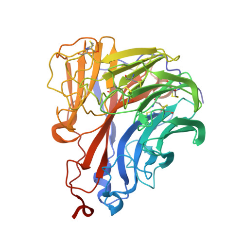
5NWE

Complex of H275Y mutant variant of neuraminidase from H1N1 influenza virus with oseltamivir


Experimental Data Snapshot

  • Method: X-RAY DIFFRACTION
  • Resolution: 2.00 Å
  • R-Value Free: 0.277 
  • R-Value Work: 0.235 
  • R-Value Observed: 0.236 

Starting Model: experimental
View more details

wwPDB Validation   3D Report Full Report


Ligand Structure Quality Assessment 


This is version 2.1 of the entry. See complete history


Literature

Kinetic, Thermodynamic, and Structural Analysis of Drug Resistance Mutations in Neuraminidase from the 2009 Pandemic Influenza Virus.

Pokorna, J.Pachl, P.Karlukova, E.Hejdanek, J.Rezacova, P.Machara, A.Hudlicky, J.Konvalinka, J.Kozisek, M.

(2018) Viruses 10

  • DOI: https://doi.org/10.3390/v10070339
  • Primary Citation of Related Structures:  
    5NWE, 5NZ4, 5NZE, 5NZF, 5NZN

  • PubMed Abstract: 

    Neuraminidase is the main target for current influenza drugs. Reduced susceptibility to oseltamivir, the most widely prescribed neuraminidase inhibitor, has been repeatedly reported. The resistance substitutions I223V and S247N, alone or in combination with the major oseltamivir-resistance mutation H275Y, have been observed in 2009 pandemic H1N1 viruses. We overexpressed and purified the ectodomain of wild-type neuraminidase from the A/California/07/2009 (H1N1) influenza virus, as well as variants containing H275Y, I223V, and S247N single mutations and H275Y/I223V and H275Y/S247N double mutations. We performed enzymological and thermodynamic analyses and structurally examined the resistance mechanism. Our results reveal that the I223V or S247N substitution alone confers only a moderate reduction in oseltamivir affinity. In contrast, the major oseltamivir resistance mutation H275Y causes a significant decrease in the enzyme’s ability to bind this drug. Combination of H275Y with an I223V or S247N mutation results in extreme impairment of oseltamivir’s inhibition potency. Our structural analyses revealed that the H275Y substitution has a major effect on the oseltamivir binding pose within the active site while the influence of other studied mutations is much less prominent. Our crystal structures also helped explain the augmenting effect on resistance of combining H275Y with both substitutions.


  • Organizational Affiliation

    The Czech Academy of Sciences, Institute of Organic Chemistry and Biochemistry, Flemingovo n. 2, 166 10 Prague 6, Czech Republic. [email protected].


Macromolecules
Find similar proteins by:  (by identity cutoff)  |  3D Structure
Entity ID: 1
MoleculeChains Sequence LengthOrganismDetailsImage
Neuraminidase
A, B, C, D
388Influenza A virusMutation(s): 0 
Gene Names: NA
EC: 3.2.1.18
UniProt
Find proteins for C3W6G3 (Influenza A virus)
Explore C3W6G3 
Go to UniProtKB:  C3W6G3
Entity Groups  
Sequence Clusters30% Identity50% Identity70% Identity90% Identity95% Identity100% Identity
UniProt GroupC3W6G3
Glycosylation
Glycosylation Sites: 3
Sequence Annotations
Expand
  • Reference Sequence
Oligosaccharides

Help

Entity ID: 2
MoleculeChains Length2D Diagram Glycosylation3D Interactions
alpha-D-mannopyranose-(1-6)-beta-D-mannopyranose-(1-4)-2-acetamido-2-deoxy-beta-D-glucopyranose-(1-4)-[alpha-L-fucopyranose-(1-6)]2-acetamido-2-deoxy-beta-D-glucopyranose
E
5N-Glycosylation
Glycosylation Resources
GlyTouCan:  G00395TQ
GlyCosmos:  G00395TQ
GlyGen:  G00395TQ
Entity ID: 3
MoleculeChains Length2D Diagram Glycosylation3D Interactions
2-acetamido-2-deoxy-beta-D-glucopyranose-(1-4)-2-acetamido-2-deoxy-beta-D-glucopyranose
F
2N-Glycosylation
Glycosylation Resources
GlyTouCan:  G42666HT
GlyCosmos:  G42666HT
GlyGen:  G42666HT
Small Molecules
Ligands 3 Unique
IDChains Name / Formula / InChI Key2D Diagram3D Interactions
G39
Query on G39

Download Ideal Coordinates CCD File 
I [auth A],
N [auth B],
T [auth C],
Y [auth D]
(3R,4R,5S)-4-(acetylamino)-5-amino-3-(pentan-3-yloxy)cyclohex-1-ene-1-carboxylic acid
C14 H24 N2 O4
NENPYTRHICXVCS-YNEHKIRRSA-N
NAG
Query on NAG

Download Ideal Coordinates CCD File 
AA [auth D]
BA [auth D]
J [auth A]
K [auth A]
O [auth B]
AA [auth D],
BA [auth D],
J [auth A],
K [auth A],
O [auth B],
P [auth B],
Q [auth B],
U [auth C],
V [auth C],
Z [auth D]
2-acetamido-2-deoxy-beta-D-glucopyranose
C8 H15 N O6
OVRNDRQMDRJTHS-FMDGEEDCSA-N
CA
Query on CA

Download Ideal Coordinates CCD File 
G [auth A]
H [auth A]
L [auth B]
M [auth B]
R [auth C]
G [auth A],
H [auth A],
L [auth B],
M [auth B],
R [auth C],
S [auth C],
W [auth D],
X [auth D]
CALCIUM ION
Ca
BHPQYMZQTOCNFJ-UHFFFAOYSA-N
Experimental Data & Validation

Experimental Data

  • Method: X-RAY DIFFRACTION
  • Resolution: 2.00 Å
  • R-Value Free: 0.277 
  • R-Value Work: 0.235 
  • R-Value Observed: 0.236 
  • Space Group: P 1 21 1
Unit Cell:
Length ( Å )Angle ( ˚ )
a = 85.474α = 90
b = 126.608β = 93.49
c = 96.56γ = 90
Software Package:
Software NamePurpose
REFMACrefinement
XDSdata scaling
XDSdata reduction
MOLREPphasing

Structure Validation

View Full Validation Report


Ligand Structure Quality Assessment 


Entry History & Funding Information

Deposition Data


Funding OrganizationLocationGrant Number
Grant Agency of the Czech RepublicCzech Republic13-19561S
Ministry of Education, Youth and Sports of the Czech RepublicCzech RepublicInterBioMed LO1302
Ministry of Education, Youth and Sports of the Czech RepublicCzech RepublicInterBioMed LO1304

Revision History  (Full details and data files)

  • Version 1.0: 2018-07-04
    Type: Initial release
  • Version 2.0: 2020-07-29
    Type: Remediation
    Reason: Carbohydrate remediation
    Changes: Advisory, Atomic model, Data collection, Derived calculations, Structure summary
  • Version 2.1: 2024-01-17
    Changes: Data collection, Database references, Derived calculations, Refinement description, Structure summary