5T67

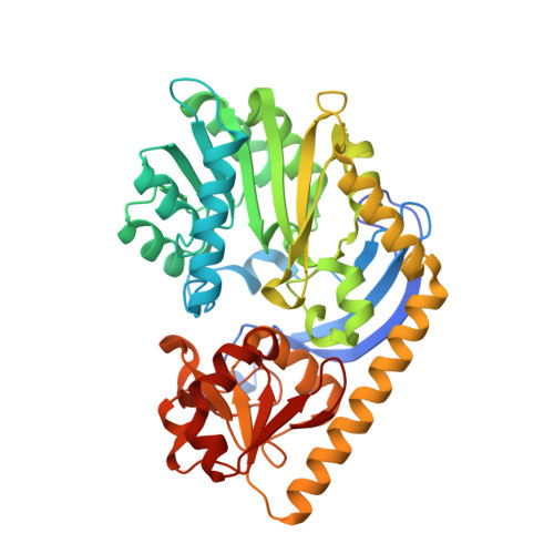
x-ray structure of the KijD1 C3-methyltransferase from Actinomadura kijaniata in complex with SAH and dTDP-sugar product


Experimental Data Snapshot

  • Method: X-RAY DIFFRACTION
  • Resolution: 1.60 Å
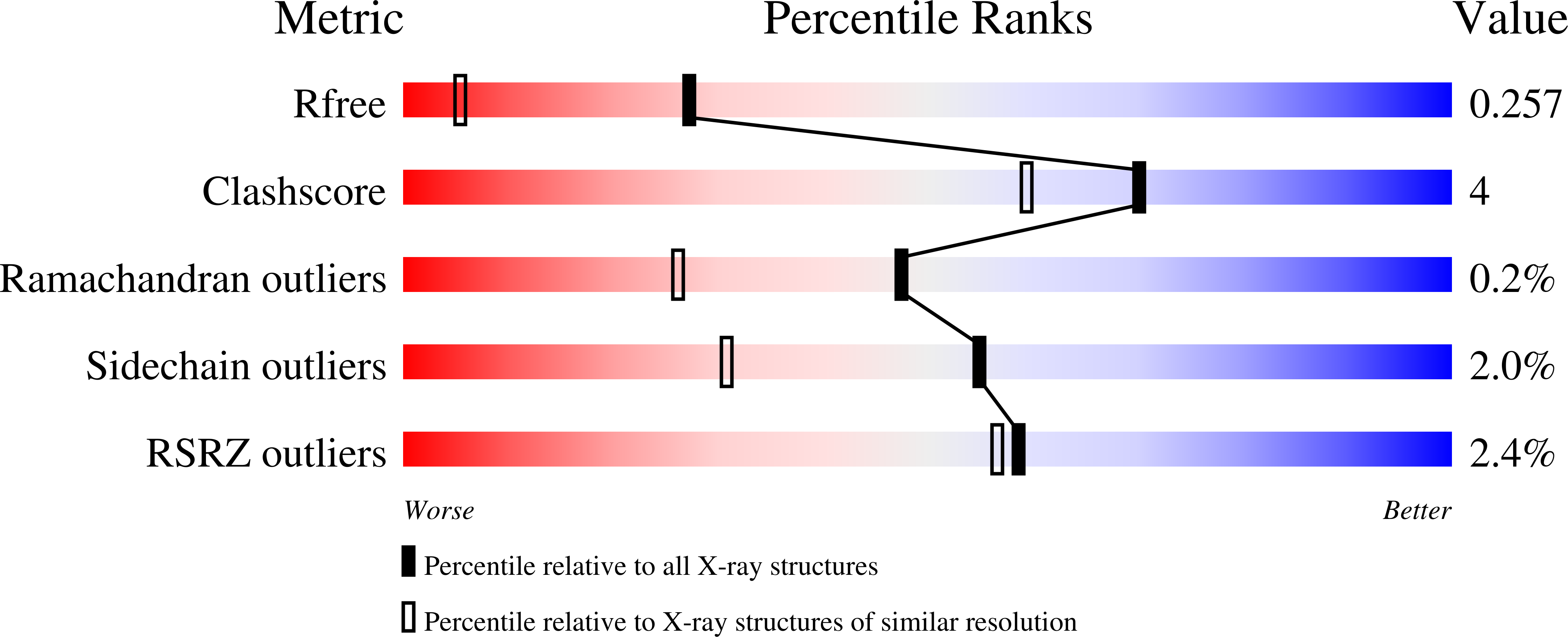
  • R-Value Free: 0.249 
  • R-Value Work: 0.198 
  • R-Value Observed: 0.201 

Starting Model: experimental
View more details

wwPDB Validation   3D Report Full Report


Ligand Structure Quality Assessment 


This is version 1.6 of the entry. See complete history


Literature

Structural studies on KijD1, a sugar C-3'-methyltransferase.

Dow, G.T.Thoden, J.B.Holden, H.M.

(2016) Protein Sci 25: 2282-2289

  • DOI: https://doi.org/10.1002/pro.3034
  • Primary Citation of Related Structures:  
    5T64, 5T67, 5T6B

  • PubMed Abstract: 

    Kijanimicin is an antitumor antibiotic isolated from Actinomadura kijaniata. It is composed of three distinct moieties: a pentacyclic core, a monosaccharide referred to as d-kijanose, and a tetrasaccharide chain composed of l-digitoxose units. d-Kijanose is a highly unusual nitro-containing tetradeoxysugar, which requires at least ten enzymes for its production. Here we describe a structural analysis of one of these enzymes, namely KijD1, which functions as a C-3'-methyltransferase using S-adenosylmethionine as its cofactor. For this investigation, two ternary complexes of KijD1, determined in the presence of S-adenosylhomocysteine (SAH) and dTDP or SAH and dTDP-3-amino-2,3,6-trideoxy-4-keto-3-methyl-d-glucose, were solved to 1.7 or 1.6 Å resolution, respectively. Unexpectedly, these structures, as well as additional biochemical analyses, demonstrated that the quaternary structure of KijD1 is a dimer. Indeed, this is in sharp contrast to that previously observed for the sugar C-3'-methyltransferase isolated from Micromonospora chalcea. By the judicious use of site-directed mutagenesis, it was possible to convert the dimeric form of KijD1 into a monomeric version. The quaternary structure of KijD1 could not have been deduced based solely on bioinformatics approaches, and thus this investigation highlights the continuing need for experimental validation.


  • Organizational Affiliation

    Department of Biochemistry, University of Wisconsin, Madison, WI, 53706.


Macromolecules
Find similar proteins by:  (by identity cutoff)  |  3D Structure
Entity ID: 1
MoleculeChains Sequence LengthOrganismDetailsImage
Sugar 3-C-methyl transferase
A, B
416Actinomadura kijaniataMutation(s): 0 
Gene Names: KijD1
UniProt
Find proteins for B3TMQ9 (Actinomadura kijaniata)
Explore B3TMQ9 
Go to UniProtKB:  B3TMQ9
Entity Groups  
Sequence Clusters30% Identity50% Identity70% Identity90% Identity95% Identity100% Identity
UniProt GroupB3TMQ9
Sequence Annotations
Expand
  • Reference Sequence
Small Molecules
Ligands 6 Unique
IDChains Name / Formula / InChI Key2D Diagram3D Interactions
JHZ
Query on JHZ

Download Ideal Coordinates CCD File 
E [auth A],
N [auth B]
(2R,4S,6R)-4-amino-4,6-dimethyl-5-oxotetrahydro-2H-pyran-2-yl [(2R,3S,5R)-3-hydroxy-5-(5-methyl-2,4-dioxo-3,4-dihydropyrimidin-1(2H)-yl)tetrahydrofuran-2-yl]methyl dihydrogen diphosphate (non-preferred name)
C17 H27 N3 O13 P2
LXNLHPYBLNFMEH-XXXNSVIPSA-N
SAH
Query on SAH

Download Ideal Coordinates CCD File 
D [auth A],
M [auth B]
S-ADENOSYL-L-HOMOCYSTEINE
C14 H20 N6 O5 S
ZJUKTBDSGOFHSH-WFMPWKQPSA-N
ZN
Query on ZN

Download Ideal Coordinates CCD File 
C [auth A],
L [auth B]
ZINC ION
Zn
PTFCDOFLOPIGGS-UHFFFAOYSA-N
EDO
Query on EDO

Download Ideal Coordinates CCD File 
F [auth A],
G [auth A],
H [auth A],
O [auth B]
1,2-ETHANEDIOL
C2 H6 O2
LYCAIKOWRPUZTN-UHFFFAOYSA-N
CL
Query on CL

Download Ideal Coordinates CCD File 
K [auth A]CHLORIDE ION
Cl
VEXZGXHMUGYJMC-UHFFFAOYSA-M
MG
Query on MG

Download Ideal Coordinates CCD File 
I [auth A],
J [auth A]
MAGNESIUM ION
Mg
JLVVSXFLKOJNIY-UHFFFAOYSA-N
Experimental Data & Validation

Experimental Data

  • Method: X-RAY DIFFRACTION
  • Resolution: 1.60 Å
  • R-Value Free: 0.249 
  • R-Value Work: 0.198 
  • R-Value Observed: 0.201 
  • Space Group: P 21 21 21
Unit Cell:
Length ( Å )Angle ( ˚ )
a = 51.088α = 90
b = 108.893β = 90
c = 142.783γ = 90
Software Package:
Software NamePurpose
REFMACrefinement
SAINTdata reduction
SADABSdata scaling
PHASERphasing

Structure Validation

View Full Validation Report


Ligand Structure Quality Assessment 


Entry History & Funding Information

Deposition Data


Funding OrganizationLocationGrant Number
National Institutes of Health/National Institute of General Medical Sciences (NIH/NIGMS)United Statesgm115921

Revision History  (Full details and data files)

  • Version 1.0: 2016-09-14
    Type: Initial release
  • Version 1.1: 2016-09-21
    Changes: Database references
  • Version 1.2: 2016-09-28
    Changes: Structure summary
  • Version 1.3: 2016-12-07
    Changes: Database references
  • Version 1.4: 2017-09-27
    Changes: Author supporting evidence, Database references
  • Version 1.5: 2019-12-25
    Changes: Author supporting evidence
  • Version 1.6: 2023-10-04
    Changes: Data collection, Database references, Derived calculations, Refinement description