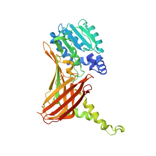
5TBI

Crystal structure of mouse CARM1 in complex with inhibitor LH1427


Experimental Data Snapshot

  • Method: X-RAY DIFFRACTION
  • Resolution: 2.29 Å
  • R-Value Free: 0.228 
  • R-Value Work: 0.198 
  • R-Value Observed: 0.199 

Starting Model: experimental
View more details

wwPDB Validation   3D Report Full Report


Ligand Structure Quality Assessment 


This is version 1.2 of the entry. See complete history


Literature

Hijacking DNA methyltransferase transition state analogues to produce chemical scaffolds for PRMT inhibitors.

Halby, L.Marechal, N.Pechalrieu, D.Cura, V.Franchini, D.M.Faux, C.Alby, F.Troffer-Charlier, N.Kudithipudi, S.Jeltsch, A.Aouadi, W.Decroly, E.Guillemot, J.C.Page, P.Ferroud, C.Bonnefond, L.Guianvarc'h, D.Cavarelli, J.Arimondo, P.B.

(2018) Philos Trans R Soc Lond B Biol Sci 373

  • DOI: https://doi.org/10.1098/rstb.2017.0072
  • Primary Citation of Related Structures:  
    5LV2, 5LV3, 5LV4, 5LV5, 5TBH, 5TBI, 5TBJ

  • PubMed Abstract: 

    DNA, RNA and histone methylation is implicated in various human diseases such as cancer or viral infections, playing a major role in cell process regulation, especially in modulation of gene expression. Here we developed a convergent synthetic pathway starting from a protected bromomethylcytosine derivative to synthesize transition state analogues of the DNA methyltransferases. This approach led to seven 5-methylcytosine-adenosine compounds that were, surprisingly, inactive against hDNMT1, hDNMT3Acat, TRDMT1 and other RNA human and viral methyltransferases. Interestingly, compound 4 and its derivative 2 showed an inhibitory activity against PRMT4 in the micromolar range. Crystal structures showed that compound 4 binds to the PRMT4 active site, displacing strongly the S -adenosyl-l-methionine cofactor, occupying its binding site, and interacting with the arginine substrate site through the cytosine moiety that probes the space filled by a substrate peptide methylation intermediate. Furthermore, the binding of the compounds induces important structural switches. These findings open new routes for the conception of new potent PRMT4 inhibitors based on the 5-methylcytosine-adenosine scaffold.This article is part of a discussion meeting issue 'Frontiers in epigenetic chemical biology'.


  • Organizational Affiliation

    CNRS FRE3600 ETaC, bât. IBCG, 31062 Toulouse, France.


Macromolecules
Find similar proteins by:  (by identity cutoff)  |  3D Structure
Entity ID: 1
MoleculeChains Sequence LengthOrganismDetailsImage
Histone-arginine methyltransferase CARM1
A, B, C, D
361Mus musculusMutation(s): 0 
Gene Names: Carm1Prmt4
EC: 2.1.1.319
UniProt & NIH Common Fund Data Resources
Find proteins for Q9WVG6 (Mus musculus)
Explore Q9WVG6 
Go to UniProtKB:  Q9WVG6
IMPC:  MGI:1913208
Entity Groups  
Sequence Clusters30% Identity50% Identity70% Identity90% Identity95% Identity100% Identity
UniProt GroupQ9WVG6
Sequence Annotations
Expand
  • Reference Sequence
Small Molecules
Ligands 4 Unique
IDChains Name / Formula / InChI Key2D Diagram3D Interactions
78V
Query on 78V

Download Ideal Coordinates CCD File 
O [auth B],
Z [auth D]
4-[2-[[(2~{R},3~{S},4~{R},5~{R})-5-(6-aminopurin-9-yl)-3,4-bis(oxidanyl)oxolan-2-yl]methylamino]ethylamino]-1-(methoxymethyl)pyrimidin-2-one
C18 H25 N9 O5
ZZOHCVFRIAWCAC-IWCJZZDYSA-N
SAO
Query on SAO

Download Ideal Coordinates CCD File 
K [auth A],
V [auth C]
5'-S-[(3S)-3-azaniumyl-3-carboxypropyl]-5'-thioadenosine
C14 H21 N6 O5 S
ZJUKTBDSGOFHSH-WFMPWKQPSA-O
PE8
Query on PE8

Download Ideal Coordinates CCD File 
L [auth B]3,6,9,12,15,18,21-HEPTAOXATRICOSANE-1,23-DIOL
C16 H34 O9
GLZWNFNQMJAZGY-UHFFFAOYSA-N
EDO
Query on EDO

Download Ideal Coordinates CCD File 
E [auth A]
F [auth A]
G [auth A]
H [auth A]
I [auth A]
E [auth A],
F [auth A],
G [auth A],
H [auth A],
I [auth A],
J [auth A],
M [auth B],
N [auth B],
P [auth C],
Q [auth C],
R [auth C],
S [auth C],
T [auth C],
U [auth C],
W [auth D],
X [auth D],
Y [auth D]
1,2-ETHANEDIOL
C2 H6 O2
LYCAIKOWRPUZTN-UHFFFAOYSA-N
Experimental Data & Validation

Experimental Data

  • Method: X-RAY DIFFRACTION
  • Resolution: 2.29 Å
  • R-Value Free: 0.228 
  • R-Value Work: 0.198 
  • R-Value Observed: 0.199 
  • Space Group: P 21 21 2
Unit Cell:
Length ( Å )Angle ( ˚ )
a = 74.494α = 90
b = 98.029β = 90
c = 205.73γ = 90
Software Package:
Software NamePurpose
PHENIXrefinement
XDSdata reduction
XDSdata scaling
PHASERphasing

Structure Validation

View Full Validation Report


Ligand Structure Quality Assessment 


Entry History 

Deposition Data

Revision History  (Full details and data files)

  • Version 1.0: 2017-09-20
    Type: Initial release
  • Version 1.1: 2019-04-03
    Changes: Data collection, Database references
  • Version 1.2: 2024-01-17
    Changes: Data collection, Database references, Refinement description