5U21

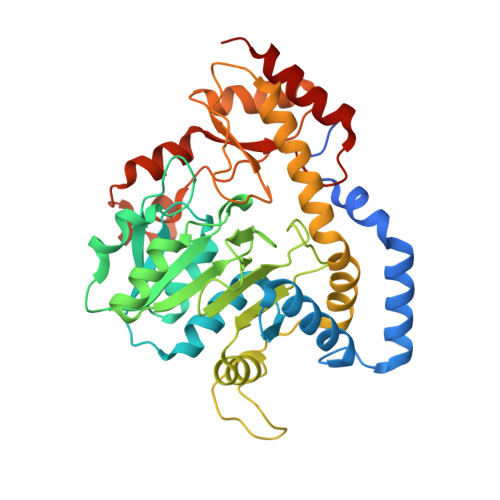
X-ray structure of the WlaRF aminotransferase from Campylobacter jejuni, K184A mutant in complex with TDP-Qui3N


Experimental Data Snapshot

  • Method: X-RAY DIFFRACTION
  • Resolution: 1.60 Å
  • R-Value Free: 0.161 
  • R-Value Work: 0.132 
  • R-Value Observed: 0.134 

Starting Model: experimental
View more details

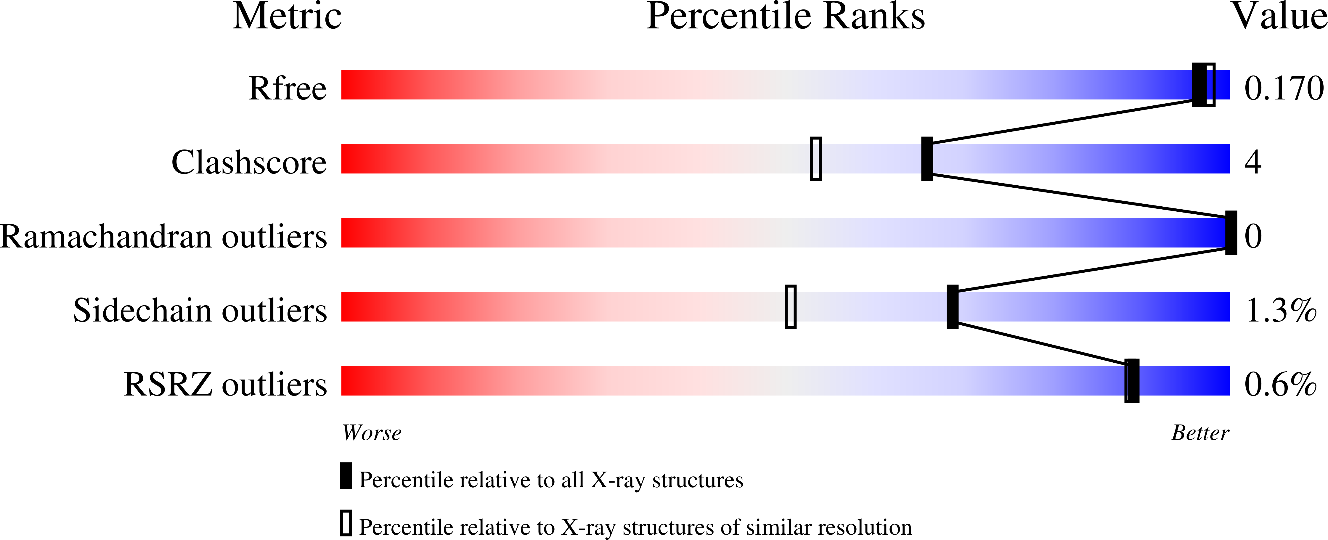
wwPDB Validation   3D Report Full Report


Ligand Structure Quality Assessment 


This is version 1.4 of the entry. See complete history


Literature

Structural investigation on WlaRG from Campylobacter jejuni: A sugar aminotransferase.

Dow, G.T.Gilbert, M.Thoden, J.B.Holden, H.M.

(2017) Protein Sci 26: 586-599

  • DOI: https://doi.org/10.1002/pro.3109
  • Primary Citation of Related Structures:  
    3NYS, 3NYT, 3NYU, 5U1Z, 5U20, 5U21, 5U23, 5U24

  • PubMed Abstract: 

    Campylobacter jejuni is a Gram-negative bacterium that represents a leading cause of human gastroenteritis worldwide. Of particular concern is the link between C. jejuni infections and the subsequent development of Guillain-Barré syndrome, an acquired autoimmune disorder leading to paralysis. All Gram-negative bacteria contain complex glycoconjugates anchored to their outer membranes, but in most strains of C. jejuni, this lipoglycan lacks the O-antigen repeating units. Recent mass spectrometry analyses indicate that the C. jejuni 81116 (Penner serotype HS:6) lipoglycan contains two dideoxyhexosamine residues, and enzymological assay data show that this bacterial strain can synthesize both dTDP-3-acetamido-3,6-dideoxy-d-glucose and dTDP-3-acetamido-3,6-dideoxy-d-galactose. The focus of this investigation is on WlaRG from C. jejuni, which plays a key role in the production of these unusual sugars by functioning as a pyridoxal 5'-phosphate dependent aminotransferase. Here, we describe the first three-dimensional structures of the enzyme in various complexes determined to resolutions of 1.7 Å or higher. Of particular significance are the external aldimine structures of WlaRG solved in the presence of either dTDP-3-amino-3,6-dideoxy-d-galactose or dTDP-3-amino-3,6-dideoxy-d-glucose. These models highlight the manner in which WlaRG can accommodate sugars with differing stereochemistries about their C-4' carbon positions. In addition, we present a corrected structure of WbpE, a related sugar aminotransferase from Pseudomonas aeruginosa, solved to 1.3 Å resolution.


  • Organizational Affiliation

    Department of Biochemistry, University of Wisconsin, Madison, Wisconsin, 53706.


Macromolecules
Find similar proteins by:  (by identity cutoff)  |  3D Structure
Entity ID: 1
MoleculeChains Sequence LengthOrganismDetailsImage
Putative aminotransferase
A, B, C, D
382Campylobacter jejuniMutation(s): 1 
UniProt
Find proteins for Q9ALS9 (Campylobacter jejuni)
Explore Q9ALS9 
Go to UniProtKB:  Q9ALS9
Entity Groups  
Sequence Clusters30% Identity50% Identity70% Identity90% Identity95% Identity100% Identity
UniProt GroupQ9ALS9
Sequence Annotations
Expand
  • Reference Sequence
Small Molecules
Ligands 4 Unique
IDChains Name / Formula / InChI Key2D Diagram3D Interactions
TQP
Query on TQP

Download Ideal Coordinates CCD File 
F [auth A],
K [auth B],
N [auth C],
O [auth D]
(2R,3R,4S,5S,6R)-3,5-dihydroxy-4-{[(1E)-{3-hydroxy-2-methyl-5-[(phosphonooxy)methyl]pyridin-4-yl}methylidene]amino}-6-methyltetrahydro-2H-pyran-2-yl [(2R,3S,5R)-3-hydroxy-5-(5-methyl-2,4-dioxo-3,4-dihydropyrimidin-1(2H)-yl)tetrahydrofuran-2-yl]methyl dihydrogen diphosphate
C24 H35 N4 O19 P3
UJDWKFAFGYHSFM-HMSDFHDVSA-N
EDO
Query on EDO

Download Ideal Coordinates CCD File 
G [auth A]
H [auth A]
I [auth A]
L [auth B]
P [auth D]
G [auth A],
H [auth A],
I [auth A],
L [auth B],
P [auth D],
Q [auth D]
1,2-ETHANEDIOL
C2 H6 O2
LYCAIKOWRPUZTN-UHFFFAOYSA-N
CL
Query on CL

Download Ideal Coordinates CCD File 
J [auth A]CHLORIDE ION
Cl
VEXZGXHMUGYJMC-UHFFFAOYSA-M
NA
Query on NA

Download Ideal Coordinates CCD File 
E [auth A],
M [auth C]
SODIUM ION
Na
FKNQFGJONOIPTF-UHFFFAOYSA-N
Experimental Data & Validation

Experimental Data

  • Method: X-RAY DIFFRACTION
  • Resolution: 1.60 Å
  • R-Value Free: 0.161 
  • R-Value Work: 0.132 
  • R-Value Observed: 0.134 
  • Space Group: P 1 21 1
Unit Cell:
Length ( Å )Angle ( ˚ )
a = 108.164α = 90
b = 56.653β = 90.04
c = 124.802γ = 90
Software Package:
Software NamePurpose
REFMACrefinement
SAINTdata reduction
SADABSdata scaling
PHASERphasing

Structure Validation

View Full Validation Report


Ligand Structure Quality Assessment 


Entry History & Funding Information

Deposition Data


Funding OrganizationLocationGrant Number
National Institutes of Health/National Institute of General Medical Sciences (NIH/NIGMS)United StatesGM115921

Revision History  (Full details and data files)

  • Version 1.0: 2017-01-11
    Type: Initial release
  • Version 1.1: 2017-03-08
    Changes: Database references
  • Version 1.2: 2017-09-27
    Changes: Author supporting evidence, Structure summary
  • Version 1.3: 2020-01-01
    Changes: Author supporting evidence
  • Version 1.4: 2023-10-04
    Changes: Data collection, Database references, Derived calculations, Refinement description