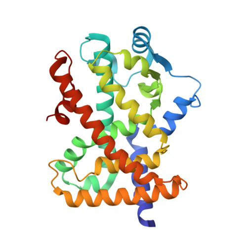
5U3Z

Human PPARdelta ligand-binding domain in complexed with specific agonist 10


Experimental Data Snapshot

  • Method: X-RAY DIFFRACTION
  • Resolution: 1.72 Å
  • R-Value Free: 0.214 
  • R-Value Work: 0.172 
  • R-Value Observed: 0.173 

Starting Model: experimental
View more details

wwPDB Validation   3D Report Full Report


Ligand Structure Quality Assessment 


This is version 2.1 of the entry. See complete history


Literature

Structural basis for specific ligation of the peroxisome proliferator-activated receptor delta.

Wu, C.C.Baiga, T.J.Downes, M.La Clair, J.J.Atkins, A.R.Richard, S.B.Fan, W.Stockley-Noel, T.A.Bowman, M.E.Noel, J.P.Evans, R.M.

(2017) Proc Natl Acad Sci U S A 114: E2563-E2570

  • DOI: https://doi.org/10.1073/pnas.1621513114
  • Primary Citation of Related Structures:  
    5U3Q, 5U3R, 5U3S, 5U3T, 5U3U, 5U3V, 5U3W, 5U3X, 5U3Y, 5U3Z, 5U40, 5U41, 5U42, 5U43, 5U44, 5U45, 5U46

  • PubMed Abstract: 

    The peroxisome proliferator-activated receptor (PPAR) family comprises three subtypes: PPARα, PPARγ, and PPARδ. PPARδ transcriptionally modulates lipid metabolism and the control of energy homeostasis; therefore, PPARδ agonists are promising agents for treating a variety of metabolic disorders. In the present study, we develop a panel of rationally designed PPARδ agonists. The modular motif affords efficient syntheses using building blocks optimized for interactions with subtype-specific residues in the PPARδ ligand-binding domain (LBD). A combination of atomic-resolution protein X-ray crystallographic structures, ligand-dependent LBD stabilization assays, and cell-based transactivation measurements delineate structure-activity relationships (SARs) for PPARδ-selective targeting and structural modulation. We identify key ligand-induced conformational transitions of a conserved tryptophan side chain in the LBD that trigger reorganization of the H2'-H3 surface segment of PPARδ. The subtype-specific conservation of H2'-H3 sequences suggests that this architectural remodeling constitutes a previously unrecognized conformational switch accompanying ligand-dependent PPARδ transcriptional regulation.


  • Organizational Affiliation

    Howard Hughes Medical Institute, The Salk Institute for Biological Studies, La Jolla, CA 92037.


Macromolecules
Find similar proteins by:  (by identity cutoff)  |  3D Structure
Entity ID: 1
MoleculeChains Sequence LengthOrganismDetailsImage
Peroxisome proliferator-activated receptor delta
A, B
272Homo sapiensMutation(s): 0 
Gene Names: PPARDNR1C2PPARB
UniProt & NIH Common Fund Data Resources
Find proteins for Q03181 (Homo sapiens)
Explore Q03181 
Go to UniProtKB:  Q03181
PHAROS:  Q03181
GTEx:  ENSG00000112033 
Entity Groups  
Sequence Clusters30% Identity50% Identity70% Identity90% Identity95% Identity100% Identity
UniProt GroupQ03181
Sequence Annotations
Expand
  • Reference Sequence
Small Molecules
Ligands 4 Unique
IDChains Name / Formula / InChI Key2D Diagram3D Interactions
7UA
Query on 7UA

Download Ideal Coordinates CCD File 
C [auth A],
M [auth B]
6-[2-({cyclopropyl[4-(furan-3-yl)benzene-1-carbonyl]amino}methyl)phenoxy]hexanoic acid
C27 H29 N O5
JXTGKWHQFWZAGE-UHFFFAOYSA-N
B7G
Query on B7G

Download Ideal Coordinates CCD File 
G [auth A],
J [auth A],
L [auth B]
heptyl beta-D-glucopyranoside
C13 H26 O6
NIDYWHLDTIVRJT-UJPOAAIJSA-N
PEG
Query on PEG

Download Ideal Coordinates CCD File 
E [auth A],
H [auth A],
N [auth B],
O [auth B],
Q [auth B]
DI(HYDROXYETHYL)ETHER
C4 H10 O3
MTHSVFCYNBDYFN-UHFFFAOYSA-N
PGO
Query on PGO

Download Ideal Coordinates CCD File 
D [auth A],
F [auth A],
I [auth A],
K [auth A],
P [auth B]
S-1,2-PROPANEDIOL
C3 H8 O2
DNIAPMSPPWPWGF-VKHMYHEASA-N
Experimental Data & Validation

Experimental Data

  • Method: X-RAY DIFFRACTION
  • Resolution: 1.72 Å
  • R-Value Free: 0.214 
  • R-Value Work: 0.172 
  • R-Value Observed: 0.173 
  • Space Group: P 1 21 1
Unit Cell:
Length ( Å )Angle ( ˚ )
a = 39.43α = 90
b = 94.75β = 97.68
c = 96.05γ = 90
Software Package:
Software NamePurpose
PHENIXrefinement
HKL-2000data reduction
HKL-2000data scaling
PHASERphasing

Structure Validation

View Full Validation Report


Ligand Structure Quality Assessment 


Entry History 

Deposition Data

Revision History  (Full details and data files)

  • Version 1.0: 2017-03-22
    Type: Initial release
  • Version 1.1: 2017-03-29
    Changes: Database references
  • Version 1.2: 2017-04-05
    Changes: Database references
  • Version 2.0: 2020-07-29
    Type: Remediation
    Reason: Carbohydrate remediation
    Changes: Atomic model, Data collection, Derived calculations, Structure summary
  • Version 2.1: 2023-10-04
    Changes: Data collection, Database references, Refinement description, Structure summary