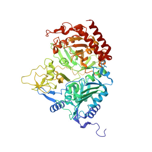
5V9F

Structure of the H477R variant of rat cytosolic PEPCK in complex with beta sulfopyruvate and GTP.

  • Classification: LYASE
  • Organism(s): Rattus norvegicus
  • Expression System: Escherichia coli
  • Mutation(s): Yes 

  • Deposited: 2017-03-23 Released: 2017-04-12 
  • Deposition Author(s): Holyoak, T., Cui, D.S.
  • Funding Organization(s): Natural Sciences and Engineering Research Council (NSERC, Canada)

Experimental Data Snapshot

  • Method: X-RAY DIFFRACTION
  • Resolution: 2.05 Å
  • R-Value Free: 0.234 
  • R-Value Work: 0.195 
  • R-Value Observed: 0.197 

Starting Model: experimental
View more details

wwPDB Validation   3D Report Full Report


Ligand Structure Quality Assessment 


This is version 1.5 of the entry. See complete history


Literature

Asymmetric Anchoring Is Required for Efficient Omega-Loop Opening and Closing in Cytosolic Phosphoenolpyruvate Carboxykinase.

Cui, D.S.Broom, A.Mcleod, M.J.Meiering, E.M.Holyoak, T.

(2017) Biochemistry 56: 2106-2115

  • DOI: https://doi.org/10.1021/acs.biochem.7b00178
  • Primary Citation of Related Structures:  
    5V95, 5V97, 5V9F, 5V9G, 5V9H

  • PubMed Abstract: 

    Mobile Ω-loops play essential roles in the function of many enzymes. Here we investigated the importance of a residue lying outside of the mobile Ω-loop element in the catalytic function of an H477R variant of cytosolic phosphoenolpyruvate carboxykinase using crystallographic, kinetic, and computational analysis. The crystallographic data suggest that the efficient transition of the Ω-loop to the closed conformation requires stabilization of the N-terminus of the loop through contacts between R461 and E588. In contrast, the C-terminal end of the Ω-loop undergoes changing interactions with the enzyme body through contacts between H477 at the C-terminus of the loop and E591 located on the enzyme body. Potential of mean force calculations demonstrated that altering the anchoring of the C-terminus of the Ω-loop via the H477R substitution results in the destabilization of the closed state of the Ω-loop by 3.4 kcal mol -1 . The kinetic parameters for the enzyme were altered in an asymmetric fashion with the predominant effect being observed in the direction of oxaloacetate synthesis. This is exemplified by a reduction in k cat for the H477R mutant by an order of magnitude in the direction of OAA synthesis, while in the direction of PEP synthesis, it decreased by a factor of only 2. The data are consistent with a mechanism for loop conformational exchange between open and closed states in which a balance between fixed anchoring of the N-terminus of the Ω-loop and a flexible, unattached C-terminus drives the transition between a disordered (open) state and an ordered (closed) state.


  • Organizational Affiliation

    Department of Biology and ‡Department of Chemistry, University of Waterloo , Waterloo, ON, Canada N2L 3G1.


Macromolecules
Find similar proteins by:  (by identity cutoff)  |  3D Structure
Entity ID: 1
MoleculeChains Sequence LengthOrganismDetailsImage
Phosphoenolpyruvate carboxykinase, cytosolic [GTP]622Rattus norvegicusMutation(s): 1 
Gene Names: Pck1
EC: 4.1.1.32 (PDB Primary Data), 2.7.11 (UniProt)
UniProt
Find proteins for P07379 (Rattus norvegicus)
Explore P07379 
Go to UniProtKB:  P07379
Entity Groups  
Sequence Clusters30% Identity50% Identity70% Identity90% Identity95% Identity100% Identity
UniProt GroupP07379
Sequence Annotations
Expand
  • Reference Sequence
Small Molecules
Ligands 4 Unique
IDChains Name / Formula / InChI Key2D Diagram3D Interactions
GTP
Query on GTP

Download Ideal Coordinates CCD File 
E [auth A]GUANOSINE-5'-TRIPHOSPHATE
C10 H16 N5 O14 P3
XKMLYUALXHKNFT-UUOKFMHZSA-N
SPV
Query on SPV

Download Ideal Coordinates CCD File 
F [auth A]SULFOPYRUVATE
C3 H4 O6 S
BUTHMSUEBYPMKJ-UHFFFAOYSA-N
MN
Query on MN

Download Ideal Coordinates CCD File 
B [auth A],
C [auth A],
G [auth A]
MANGANESE (II) ION
Mn
WAEMQWOKJMHJLA-UHFFFAOYSA-N
NA
Query on NA

Download Ideal Coordinates CCD File 
D [auth A]SODIUM ION
Na
FKNQFGJONOIPTF-UHFFFAOYSA-N
Experimental Data & Validation

Experimental Data

  • Method: X-RAY DIFFRACTION
  • Resolution: 2.05 Å
  • R-Value Free: 0.234 
  • R-Value Work: 0.195 
  • R-Value Observed: 0.197 
  • Space Group: P 1 21 1
Unit Cell:
Length ( Å )Angle ( ˚ )
a = 46.285α = 90
b = 119.506β = 107.81
c = 60.707γ = 90
Software Package:
Software NamePurpose
HKL-2000data collection
HKL-2000data scaling
MOLREPphasing
REFMACrefinement
PDB_EXTRACTdata extraction
HKL-2000data reduction
DENZOdata reduction
SCALEPACKdata scaling

Structure Validation

View Full Validation Report


Ligand Structure Quality Assessment 


Entry History & Funding Information

Deposition Data


Funding OrganizationLocationGrant Number
Natural Sciences and Engineering Research Council (NSERC, Canada)Canada--

Revision History  (Full details and data files)

  • Version 1.0: 2017-04-12
    Type: Initial release
  • Version 1.1: 2017-04-26
    Changes: Database references
  • Version 1.2: 2017-09-20
    Changes: Author supporting evidence, Refinement description
  • Version 1.3: 2017-09-27
    Changes: Data collection
  • Version 1.4: 2020-01-08
    Changes: Author supporting evidence
  • Version 1.5: 2023-10-04
    Changes: Data collection, Database references, Derived calculations, Refinement description