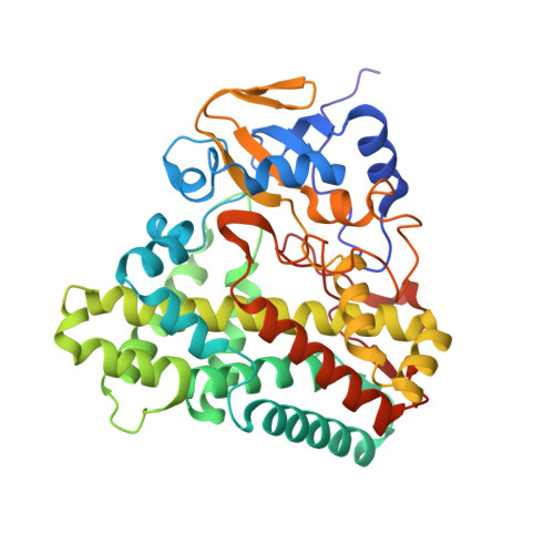
5WP2

1.44 Angstrom crystal structure of CYP121 from Mycobacterium tuberculosis in complex with substrate and CN


Experimental Data Snapshot

  • Method: X-RAY DIFFRACTION
  • Resolution: 1.44 Å
  • R-Value Free: 0.195 
  • R-Value Work: 0.179 
  • R-Value Observed: 0.180 

Starting Model: experimental
View more details

wwPDB Validation   3D Report Full Report


Ligand Structure Quality Assessment 


This is version 1.2 of the entry. See complete history


Literature

Probing Ligand Exchange in the P450 Enzyme CYP121 from Mycobacterium tuberculosis: Dynamic Equilibrium of the Distal Heme Ligand as a Function of pH and Temperature.

Fielding, A.J.Dornevil, K.Ma, L.Davis, I.Liu, A.

(2017) J Am Chem Soc 139: 17484-17499

  • DOI: https://doi.org/10.1021/jacs.7b08911
  • Primary Citation of Related Structures:  
    5WP2

  • PubMed Abstract: 

    CYP121 is a cytochrome P450 enzyme from Mycobacterium tuberculosis that catalyzes the formation of a C-C bond between the aromatic groups of its cyclodityrosine substrate (cYY). The crystal structure of CYP121 in complex with cYY reveals that the solvent-derived ligand remains bound to the ferric ion in the enzyme-substrate complex. Whereas in the generally accepted P450 mechanism, binding of the primary substrate in the active-site triggers the release of the solvent-derived ligand, priming the metal center for reduction and subsequent O 2 binding. Here we employed sodium cyanide to probe the metal-ligand exchange of the enzyme and the enzyme-substrate complex. The cyano adducts were characterized by UV-vis, EPR, and ENDOR spectroscopies and X-ray crystallography. A 100-fold increase in the affinity of cyanide binding to the enzyme-substrate complex over the ligand-free enzyme was observed. The crystal structure of the [CYP121(cYY)CN] ternary complex showed a rearrangement of the substrate in the active-site, when compared to the structure of the binary [CYP121(cYY)] complex. Transient kinetic studies showed that cYY binding resulted in a lower second-order rate constant (k on (CN) ) but a much more stable cyanide adduct with 3 orders of magnitude slower k off (CN) rate. A dynamic equilibrium between multiple high- and low-spin species for both the enzyme and enzyme-substrate complex was also observed, which is sensitive to changes in both pH and temperature. Our data reveal the chemical and physical properties of the solvent-derived ligand of the enzyme, which will help to understand the initial steps of the catalytic mechanism.


  • Organizational Affiliation

    Department of Chemistry, University of Texas at San Antonio , San Antonio, Texas 78249, United States.


Macromolecules
Find similar proteins by:  (by identity cutoff)  |  3D Structure
Entity ID: 1
MoleculeChains Sequence LengthOrganismDetailsImage
Mycocyclosin synthase395Mycobacterium tuberculosisMutation(s): 0 
Gene Names: cyp121MT2336
EC: 1.14.21.9 (PDB Primary Data), 1.14.19.70 (UniProt)
UniProt
Find proteins for P9WPP7 (Mycobacterium tuberculosis (strain ATCC 25618 / H37Rv))
Explore P9WPP7 
Go to UniProtKB:  P9WPP7
Entity Groups  
Sequence Clusters30% Identity50% Identity70% Identity90% Identity95% Identity100% Identity
UniProt GroupP9WPP7
Sequence Annotations
Expand
  • Reference Sequence
Small Molecules
Ligands 4 Unique
IDChains Name / Formula / InChI Key2D Diagram3D Interactions
HEM
Query on HEM

Download Ideal Coordinates CCD File 
B [auth A]PROTOPORPHYRIN IX CONTAINING FE
C34 H32 Fe N4 O4
KABFMIBPWCXCRK-RGGAHWMASA-L
YTT
Query on YTT

Download Ideal Coordinates CCD File 
C [auth A](3S,6S)-3,6-bis(4-hydroxybenzyl)piperazine-2,5-dione
C18 H18 N2 O4
NGPCLOGFGKJCBP-HOTGVXAUSA-N
SO4
Query on SO4

Download Ideal Coordinates CCD File 
E [auth A],
F [auth A],
G [auth A],
H [auth A]
SULFATE ION
O4 S
QAOWNCQODCNURD-UHFFFAOYSA-L
CYN
Query on CYN

Download Ideal Coordinates CCD File 
D [auth A]CYANIDE ION
C N
XFXPMWWXUTWYJX-UHFFFAOYSA-N
Experimental Data & Validation

Experimental Data

  • Method: X-RAY DIFFRACTION
  • Resolution: 1.44 Å
  • R-Value Free: 0.195 
  • R-Value Work: 0.179 
  • R-Value Observed: 0.180 
  • Space Group: P 65 2 2
Unit Cell:
Length ( Å )Angle ( ˚ )
a = 77.856α = 90
b = 77.856β = 90
c = 263.615γ = 120
Software Package:
Software NamePurpose
PHENIXrefinement
DENZOdata collection
SCALEPACKdata scaling
PDB_EXTRACTdata extraction
DENZOdata reduction
PHENIXphasing

Structure Validation

View Full Validation Report


Ligand Structure Quality Assessment 


Entry History & Funding Information

Deposition Data


Funding OrganizationLocationGrant Number
National Institutes of Health/National Institute of General Medical Sciences (NIH/NIGMS)United StatesGM108988
National Institutes of Health/National Institute of General Medical Sciences (NIH/NIGMS)United StatesGM107529

Revision History  (Full details and data files)

  • Version 1.0: 2018-05-30
    Type: Initial release
  • Version 1.1: 2020-01-01
    Changes: Author supporting evidence
  • Version 1.2: 2023-10-04
    Changes: Data collection, Database references, Refinement description, Structure summary