5XD5

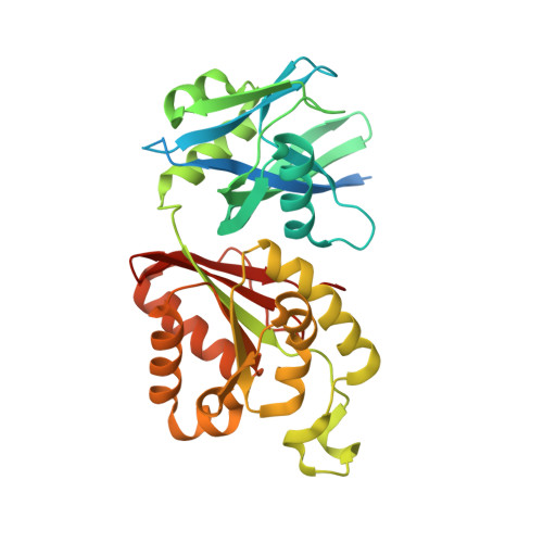
Crystal structure of Mycobacterium smegmatis MutT1 in complex with ATP, magnesium fluoride and phosphate


Experimental Data Snapshot

  • Method: X-RAY DIFFRACTION
  • Resolution: 1.75 Å
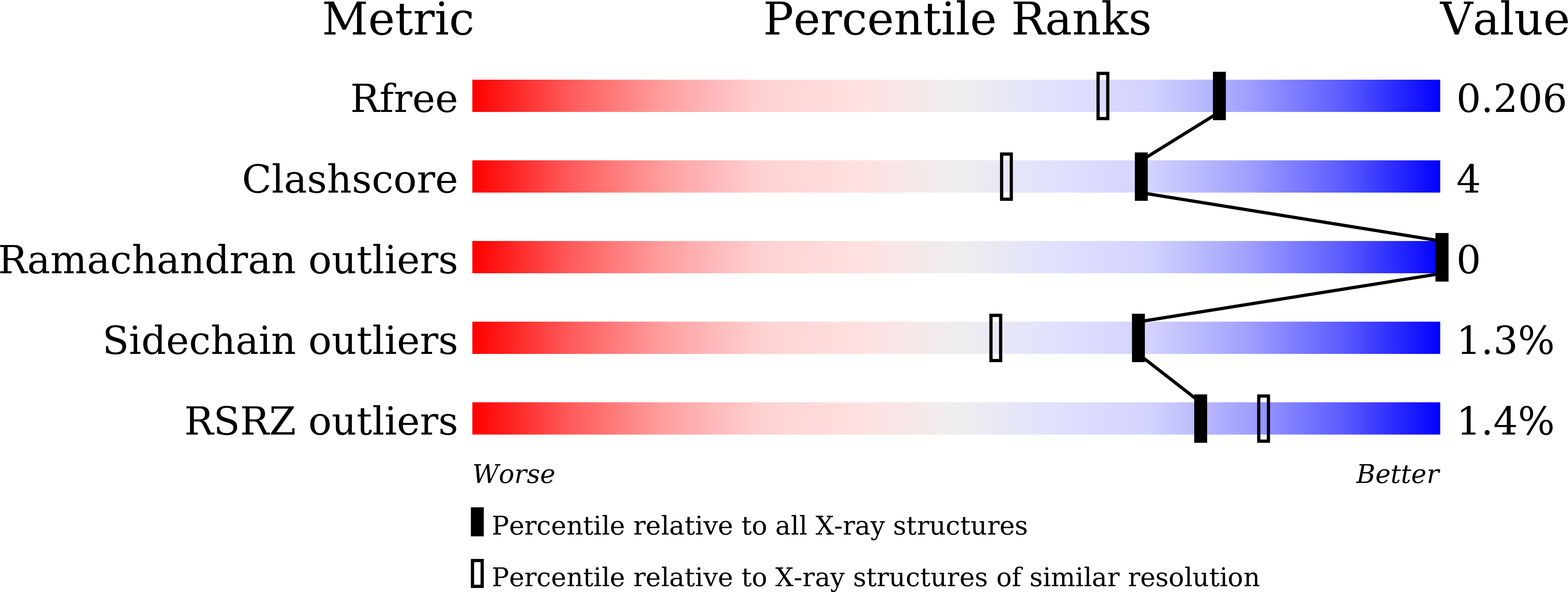
  • R-Value Free: 0.205 
  • R-Value Work: 0.166 
  • R-Value Observed: 0.168 

Starting Model: experimental
View more details

wwPDB Validation   3D Report Full Report


Ligand Structure Quality Assessment 


This is version 1.2 of the entry. See complete history


Literature

Hydrolysis of diadenosine polyphosphates. Exploration of an additional role of Mycobacterium smegmatis MutT1

Arif, S.M.Varshney, U.Vijayan, M.

(2017) J Struct Biol 199: 165-176

  • DOI: https://doi.org/10.1016/j.jsb.2017.07.002
  • Primary Citation of Related Structures:  
    5XD1, 5XD2, 5XD3, 5XD4, 5XD5

  • PubMed Abstract: 

    Diadenosine polyphosphates (Ap n A, n=2-6), particularly Ap 4 A, are involved in several important physiological processes. The substantial sequence identity of the Nudix hydrolase domain (domain 1) of Mycobacterium smegmatis MutT1 (MsMutT1) with a known Ap 4 A hydrolase suggested that MsMutT1 could also hydrolyse diadenosine polyphosphates. Biochemical experiments yielded results in conformity with this suggestion, with Ap 4 A as the best among the substrates. ATP is a product in all experiments; small amounts of ADP were also observed in the experiments involving Ap 4 A and Ap 6 A. Hydrolysis was inhibited by fluoride ions in all cases. The mechanism of action and its inhibition in relation to Ap n A were explored through the X-ray analysis of the crystals of the MsMutT1 complexes with Ap 5 A; Ap 5 A and MnCl 2 ; Ap 4 A; ATP; and ATP.NaF.MgCl 2 . The aggregation pattern of molecules in the first four crystals is similar to that found in a majority of MsMutT1-NTP crystals. Substrate molecules occupy the primary binding site and ATP occupies a site at an intermolecular interface, in the first two. ATP occupies both the sites in the third and fourth crystal. The protein-ligand interactions observed in these crystal structures lead to an explanation of the molecular mechanism of hydrolysis of Ap n A by MsMutT1. The fifth crystal exhibits a new packing arrangement. The structure of the complex provides an explanation for the fluoride inhibition of the activity of the enzyme. It would thus appear that MutT1 has a major role involving the hydrolysis of diadenosine polyphosphates, which could be elucidated at the molecular level.


  • Organizational Affiliation

    Molecular Biophysics Unit, Indian Institute of Science, Bangalore 560 012, India.


Macromolecules
Find similar proteins by:  (by identity cutoff)  |  3D Structure
Entity ID: 1
MoleculeChains Sequence LengthOrganismDetailsImage
Hydrolase, NUDIX family proteinA [auth B],
B [auth A]
342Mycolicibacterium smegmatis MC2 155Mutation(s): 0 
Gene Names: MSMEG_2390
EC: 3.6.1.61 (UniProt), 3.6.1.69 (UniProt), 3.6.1.58 (UniProt)
UniProt
Find proteins for A0QUZ2 (Mycolicibacterium smegmatis (strain ATCC 700084 / mc(2)155))
Explore A0QUZ2 
Go to UniProtKB:  A0QUZ2
Entity Groups  
Sequence Clusters30% Identity50% Identity70% Identity90% Identity95% Identity100% Identity
UniProt GroupA0QUZ2
Sequence Annotations
Expand
  • Reference Sequence
Small Molecules
Ligands 6 Unique
IDChains Name / Formula / InChI Key2D Diagram3D Interactions
ATP
Query on ATP

Download Ideal Coordinates CCD File 
C [auth B],
S [auth A]
ADENOSINE-5'-TRIPHOSPHATE
C10 H16 N5 O13 P3
ZKHQWZAMYRWXGA-KQYNXXCUSA-N
PO4
Query on PO4

Download Ideal Coordinates CCD File 
D [auth B]PHOSPHATE ION
O4 P
NBIIXXVUZAFLBC-UHFFFAOYSA-K
GOL
Query on GOL

Download Ideal Coordinates CCD File 
E [auth B]
F [auth B]
G [auth B]
H [auth B]
T [auth A]
E [auth B],
F [auth B],
G [auth B],
H [auth B],
T [auth A],
U [auth A],
V [auth A]
GLYCEROL
C3 H8 O3
PEDCQBHIVMGVHV-UHFFFAOYSA-N
CL
Query on CL

Download Ideal Coordinates CCD File 
DA [auth A]
EA [auth A]
FA [auth A]
GA [auth A]
P [auth B]
DA [auth A],
EA [auth A],
FA [auth A],
GA [auth A],
P [auth B],
Q [auth B],
R [auth B]
CHLORIDE ION
Cl
VEXZGXHMUGYJMC-UHFFFAOYSA-M
MG
Query on MG

Download Ideal Coordinates CCD File 
I [auth B]
J [auth B]
K [auth B]
L [auth B]
W [auth A]
I [auth B],
J [auth B],
K [auth B],
L [auth B],
W [auth A],
X [auth A],
Y [auth A],
Z [auth A]
MAGNESIUM ION
Mg
JLVVSXFLKOJNIY-UHFFFAOYSA-N
F
Query on F

Download Ideal Coordinates CCD File 
AA [auth A]
BA [auth A]
CA [auth A]
M [auth B]
N [auth B]
AA [auth A],
BA [auth A],
CA [auth A],
M [auth B],
N [auth B],
O [auth B]
FLUORIDE ION
F
KRHYYFGTRYWZRS-UHFFFAOYSA-M
Experimental Data & Validation

Experimental Data

  • Method: X-RAY DIFFRACTION
  • Resolution: 1.75 Å
  • R-Value Free: 0.205 
  • R-Value Work: 0.166 
  • R-Value Observed: 0.168 
  • Space Group: P 1 21 1
Unit Cell:
Length ( Å )Angle ( ˚ )
a = 50.05α = 90
b = 64.44β = 102.59
c = 103.04γ = 90
Software Package:
Software NamePurpose
REFMACrefinement
iMOSFLMdata processing
SCALAdata scaling
PHASERphasing
iMOSFLMdata reduction

Structure Validation

View Full Validation Report


Ligand Structure Quality Assessment 


Entry History 

Deposition Data

Revision History  (Full details and data files)

  • Version 1.0: 2017-08-09
    Type: Initial release
  • Version 1.1: 2017-09-27
    Changes: Database references
  • Version 1.2: 2023-11-22
    Changes: Data collection, Database references, Derived calculations, Refinement description