5XIS

Crystal structure of RNF168 UDM1 in complex with Lys63-linked diubiquitin, form I


Experimental Data Snapshot

  • Method: X-RAY DIFFRACTION
  • Resolution: 1.78 Å
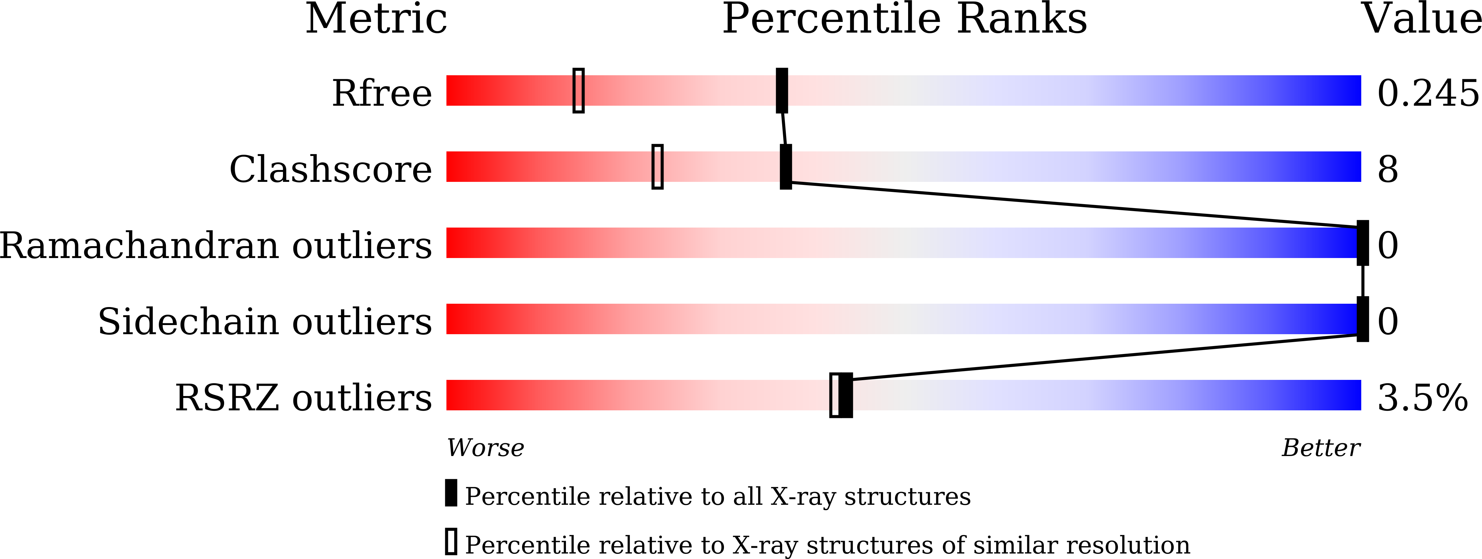
  • R-Value Free: 0.245 
  • R-Value Work: 0.218 
  • R-Value Observed: 0.220 

Starting Model: experimental
View more details

wwPDB Validation   3D Report Full Report


Ligand Structure Quality Assessment 


This is version 2.1 of the entry. See complete history


Literature

Structural insights into two distinct binding modules for Lys63-linked polyubiquitin chains in RNF168.

Takahashi, T.S.Hirade, Y.Toma, A.Sato, Y.Yamagata, A.Goto-Ito, S.Tomita, A.Nakada, S.Fukai, S.

(2018) Nat Commun 9: 170-170

  • DOI: https://doi.org/10.1038/s41467-017-02345-y
  • Primary Citation of Related Structures:  
    5XIS, 5XIT, 5XIU, 5YDK

  • PubMed Abstract: 

    The E3 ubiquitin (Ub) ligase RNF168 plays a critical role in the initiation of the DNA damage response to double-strand breaks (DSBs). The recruitment of RNF168 by ubiquitylated targets involves two distinct regions, Ub-dependent DSB recruitment module (UDM) 1 and UDM2. Here we report the crystal structures of the complex between UDM1 and Lys63-linked diUb (K63-Ub 2 ) and that between the C-terminally truncated UDM2 (UDM2ΔC) and K63-Ub 2 . In both structures, UDM1 and UDM2ΔC fold as a single α-helix. Their simultaneous bindings to the distal and proximal Ub moieties provide specificity for Lys63-linked Ub chains. Structural and biochemical analyses of UDM1 elucidate an Ub-binding mechanism between UDM1 and polyubiquitylated targets. Mutations of Ub-interacting residues in UDM2 prevent the accumulation of RNF168 to DSB sites in U2OS cells, whereas those in UDM1 have little effect, suggesting that the interaction of UDM2 with ubiquitylated and polyubiquitylated targets mainly contributes to the RNF168 recruitment.


  • Organizational Affiliation

    Institute of Molecular and Cellular Biosciences, The University of Tokyo, Tokyo, 113-0032, Japan.


Macromolecules
Find similar proteins by:  (by identity cutoff)  |  3D Structure
Entity ID: 1
MoleculeChains Sequence LengthOrganismDetailsImage
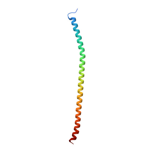
E3 ubiquitin-protein ligase RNF168
A, D
84Homo sapiensMutation(s): 0 
Gene Names: RNF168
EC: 2.3.2.27
UniProt & NIH Common Fund Data Resources
Find proteins for Q8IYW5 (Homo sapiens)
Explore Q8IYW5 
Go to UniProtKB:  Q8IYW5
PHAROS:  Q8IYW5
GTEx:  ENSG00000163961 
Entity Groups  
Sequence Clusters30% Identity50% Identity70% Identity90% Identity95% Identity100% Identity
UniProt GroupQ8IYW5
Sequence Annotations
Expand
  • Reference Sequence
Find similar proteins by:  (by identity cutoff)  |  3D Structure
Entity ID: 2
MoleculeChains Sequence LengthOrganismDetailsImage
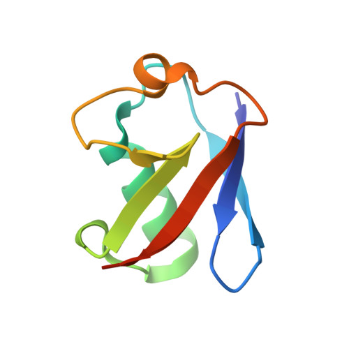
Ubiquitin-40S ribosomal protein S27a
B, E
77Mus musculusMutation(s): 0 
Gene Names: Rps27aUba80Ubcep1
UniProt
Find proteins for P62983 (Mus musculus)
Explore P62983 
Go to UniProtKB:  P62983
Entity Groups  
Sequence Clusters30% Identity50% Identity70% Identity90% Identity95% Identity100% Identity
UniProt GroupP62983
Sequence Annotations
Expand
  • Reference Sequence
Find similar proteins by:  (by identity cutoff)  |  3D Structure
Entity ID: 3
MoleculeChains Sequence LengthOrganismDetailsImage
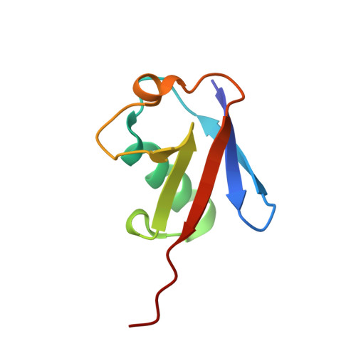
Ubiquitin-40S ribosomal protein S27a
C, F
76Mus musculusMutation(s): 1 
Gene Names: Rps27aUba80Ubcep1
UniProt
Find proteins for P62983 (Mus musculus)
Explore P62983 
Go to UniProtKB:  P62983
Entity Groups  
Sequence Clusters30% Identity50% Identity70% Identity90% Identity95% Identity100% Identity
UniProt GroupP62983
Sequence Annotations
Expand
  • Reference Sequence
Experimental Data & Validation

Experimental Data

  • Method: X-RAY DIFFRACTION
  • Resolution: 1.78 Å
  • R-Value Free: 0.245 
  • R-Value Work: 0.218 
  • R-Value Observed: 0.220 
  • Space Group: P 1
Unit Cell:
Length ( Å )Angle ( ˚ )
a = 35.273α = 76.47
b = 66.291β = 79.29
c = 74.173γ = 80.49
Software Package:
Software NamePurpose
PHENIXrefinement
HKL-2000data reduction
HKL-2000data scaling
MOLREPphasing

Structure Validation

View Full Validation Report


Ligand Structure Quality Assessment 


Entry History & Funding Information

Deposition Data


Funding OrganizationLocationGrant Number
JSPSJapan22121003
JSPSJapan15H01175
JSPSJapan24247014
JSPSJapan24687012
JSPSJapan16H04750
JSTJapanJPMJCR1XMX5
JSPSJapan15F15386

Revision History  (Full details and data files)

  • Version 1.0: 2018-03-07
    Type: Initial release
  • Version 1.1: 2018-03-28
    Changes: Structure summary
  • Version 2.0: 2020-07-29
    Type: Remediation
    Reason: Carbohydrate remediation
    Changes: Atomic model, Data collection, Derived calculations, Structure summary
  • Version 2.1: 2023-11-22
    Changes: Data collection, Database references, Refinement description, Structure summary