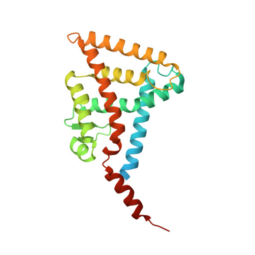
5ZQU

Crystal structure of tetrameric RXRalpha-LBD complexed with partial agonist CBt-PMN


Experimental Data Snapshot

  • Method: X-RAY DIFFRACTION
  • Resolution: 2.60 Å
  • R-Value Free: 0.270 
  • R-Value Work: 0.222 
  • R-Value Observed: 0.224 

Starting Model: experimental
View more details

wwPDB Validation   3D Report Full Report


Ligand Structure Quality Assessment 


This is version 1.2 of the entry. See complete history


Literature

Dual conformation of the ligand induces the partial agonistic activity of retinoid X receptor alpha (RXR alpha ).

Miyashita, Y.Numoto, N.Arulmozhiraja, S.Nakano, S.Matsuo, N.Shimizu, K.Shibahara, O.Fujihara, M.Kakuta, H.Ito, S.Ikura, T.Ito, N.Tokiwa, H.

(2019) FEBS Lett 593: 242-250

  • DOI: https://doi.org/10.1002/1873-3468.13301
  • Primary Citation of Related Structures:  
    5ZQU

  • PubMed Abstract: 

    1-[(3,5,5,8,8-pentamethyl-5,6,7,8-tetrahydronaphthalen-2-yl)amino]benzotriazole-5-carboxylic acid (CBt-PMN), a partial agonist of retinoid X receptor (RXR), has attracted attention due to its potential to treat type 2 diabetes and central nervous system diseases with reduced adverse effects of existing full agonists. Herein, we report the crystal structure of CBt-PMN-bound ligand-binding domain of human RXRα (hRXRα) and its biochemical characterization. Interestingly, the structure is a tetramer in nature, in which CBt-PMNs are clearly found binding in two different conformations. The dynamics of the hRXRα/CBt-PMN complex examined using molecular dynamics simulations suggest that the flexibility of the AF-2 interface depends on the conformation of the ligand. These facts reveal that the dual conformation of CBt-PMN in the complex is probably the reason behind its partial agonistic activity.


  • Organizational Affiliation

    Department of Chemistry, Rikkyo University, Tokyo, Japan.


Macromolecules
Find similar proteins by:  (by identity cutoff)  |  3D Structure
Entity ID: 1
MoleculeChains Sequence LengthOrganismDetailsImage
Retinoic acid receptor RXR-alpha
A, B, C, D
243Homo sapiensMutation(s): 0 
Gene Names: RXRANR2B1
UniProt & NIH Common Fund Data Resources
Find proteins for P19793 (Homo sapiens)
Explore P19793 
Go to UniProtKB:  P19793
PHAROS:  P19793
GTEx:  ENSG00000186350 
Entity Groups  
Sequence Clusters30% Identity50% Identity70% Identity90% Identity95% Identity100% Identity
UniProt GroupP19793
Sequence Annotations
Expand
  • Reference Sequence
Small Molecules
Ligands 2 Unique
IDChains Name / Formula / InChI Key2D Diagram3D Interactions
9HF (Subject of Investigation/LOI)
Query on 9HF

Download Ideal Coordinates CCD File 
E [auth A]
F [auth A]
K [auth B]
O [auth C]
R [auth D]
E [auth A],
F [auth A],
K [auth B],
O [auth C],
R [auth D],
S [auth D]
1-(3,5,5,8,8-pentamethyl-6,7-dihydronaphthalen-2-yl)benzotriazole-5-carboxylic acid
C22 H25 N3 O2
QSRQAKVNYDAVIT-UHFFFAOYSA-N
BR
Query on BR

Download Ideal Coordinates CCD File 
G [auth A]
H [auth A]
I [auth A]
J [auth A]
L [auth B]
G [auth A],
H [auth A],
I [auth A],
J [auth A],
L [auth B],
M [auth B],
N [auth B],
P [auth C],
Q [auth C],
T [auth D],
U [auth D]
BROMIDE ION
Br
CPELXLSAUQHCOX-UHFFFAOYSA-M
Experimental Data & Validation

Experimental Data

  • Method: X-RAY DIFFRACTION
  • Resolution: 2.60 Å
  • R-Value Free: 0.270 
  • R-Value Work: 0.222 
  • R-Value Observed: 0.224 
  • Space Group: P 1 21 1
Unit Cell:
Length ( Å )Angle ( ˚ )
a = 46.571α = 90
b = 100.492β = 99.269
c = 111.01γ = 90
Software Package:
Software NamePurpose
PHENIXrefinement
XDSdata reduction
XDSdata scaling
PHASERphasing

Structure Validation

View Full Validation Report


Ligand Structure Quality Assessment 


Entry History & Funding Information

Deposition Data


Funding OrganizationLocationGrant Number
Japan Agency for Medical Research and Development (AMED)Japan--

Revision History  (Full details and data files)

  • Version 1.0: 2019-02-27
    Type: Initial release
  • Version 1.1: 2023-11-22
    Changes: Data collection, Database references, Refinement description
  • Version 1.2: 2024-10-16
    Changes: Structure summary