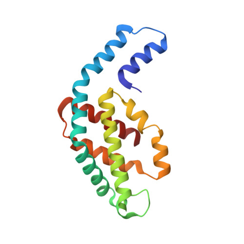
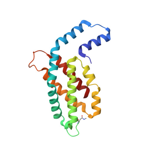
Pink-beam serial crystallography.
Meents, A., Wiedorn, M.O., Srajer, V., Henning, R., Sarrou, I., Bergtholdt, J., Barthelmess, M., Reinke, P.Y.A., Dierksmeyer, D., Tolstikova, A., Schaible, S., Messerschmidt, M., Ogata, C.M., Kissick, D.J., Taft, M.H., Manstein, D.J., Lieske, J., Oberthuer, D., Fischetti, R.F., Chapman, H.N.(2017) Nat Commun 8: 1281-1281
- PubMed: 29097720
- DOI: https://doi.org/10.1038/s41467-017-01417-3
- Primary Citation of Related Structures:
5MJL, 5MJM, 5MJP, 5MJQ, 5O7M - PubMed Abstract:
Serial X-ray crystallography allows macromolecular structure determination at both X-ray free electron lasers (XFELs) and, more recently, synchrotron sources. The time resolution for serial synchrotron crystallography experiments has been limited to millisecond timescales with monochromatic beams. The polychromatic, "pink", beam provides a more than two orders of magnitude increased photon flux and hence allows accessing much shorter timescales in diffraction experiments at synchrotron sources. Here we report the structure determination of two different protein samples by merging pink-beam diffraction patterns from many crystals, each collected with a single 100 ps X-ray pulse exposure per crystal using a setup optimized for very low scattering background. In contrast to experiments with monochromatic radiation, data from only 50 crystals were required to obtain complete datasets. The high quality of the diffraction data highlights the potential of this method for studying irreversible reactions at sub-microsecond timescales using high-brightness X-ray facilities.
Organizational Affiliation:
Center for Free Electron Laser Science, DESY, Notkestrasse 85, 22607, Hamburg, Germany. [email protected].


