5OIZ

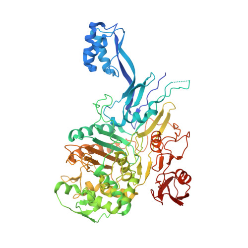
Penicillin-Binding Protein 2X (PBP2X) from Streptococcus pneumoniae in complex with oxacillin


Experimental Data Snapshot

  • Method: X-RAY DIFFRACTION
  • Resolution: 2.70 Å
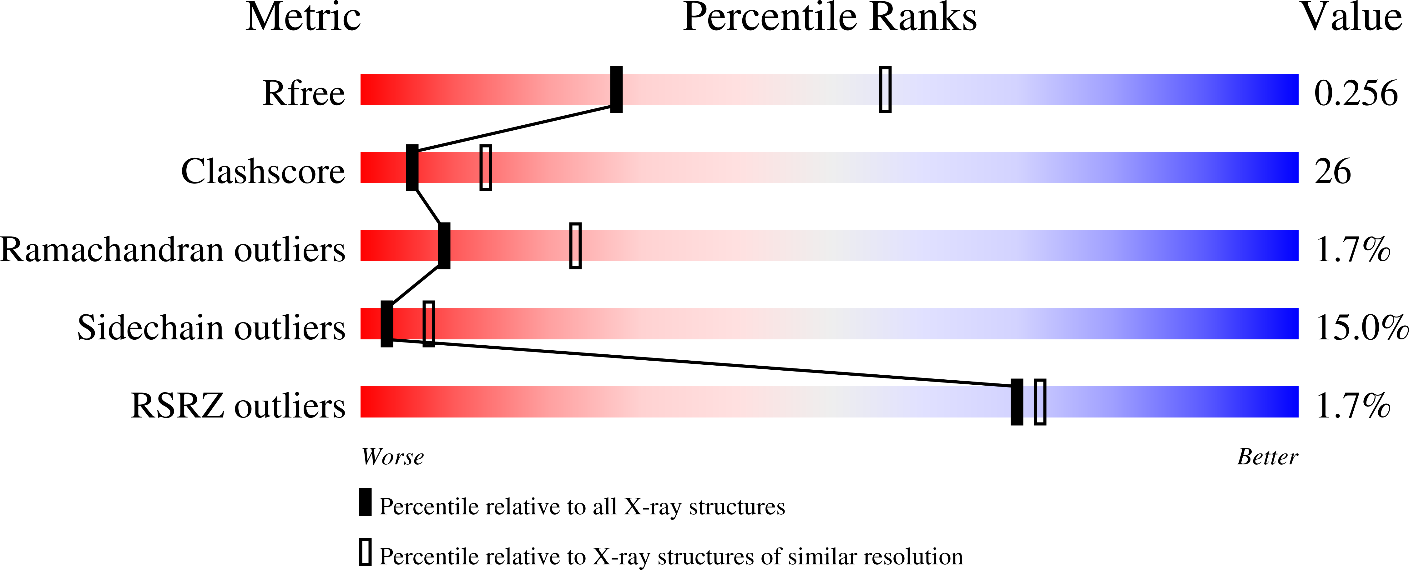
  • R-Value Free: 0.281 
  • R-Value Work: 0.217 
  • R-Value Observed: 0.220 

Starting Model: experimental
View more details

wwPDB Validation   3D Report Full Report


Ligand Structure Quality Assessment 


This is version 1.2 of the entry. See complete history


Literature

Allostery, Recognition of Nascent Peptidoglycan, and Cross-linking of the Cell Wall by the Essential Penicillin-Binding Protein 2x of Streptococcus pneumoniae.

Bernardo-Garcia, N.Mahasenan, K.V.Batuecas, M.T.Lee, M.Hesek, D.Petrackova, D.Doubravova, L.Branny, P.Mobashery, S.Hermoso, J.A.

(2018) ACS Chem Biol 13: 694-702

  • DOI: https://doi.org/10.1021/acschembio.7b00817
  • Primary Citation of Related Structures:  
    5OAU, 5OIZ, 5OJ0, 5OJ1

  • PubMed Abstract: 

    Transpeptidases, members of the penicillin-binding protein (PBP) families, catalyze cross-linking of the bacterial cell wall. This transformation is critical for the survival of bacteria, and it is the target of inhibition by β-lactam antibiotics. We report herein our structural insights into catalysis by the essential PBP2x of Streptococcus pneumoniae by disclosing a total of four X-ray structures, two computational models based on the crystal structures, and molecular-dynamics simulations. The X-ray structures are for the apo PBP2x, the enzyme modified covalently in the active site by oxacillin (a penicillin antibiotic), the enzyme modified by oxacillin in the presence of a synthetic tetrasaccharide surrogate for the cell-wall peptidoglycan, and a noncovalent complex of cefepime (a cephalosporin antibiotic) bound to the active site. A prerequisite for catalysis by transpeptidases, including PBP2x, is the molecular recognition of nascent peptidoglycan strands, which harbor pentapeptide stems. We disclose that the recognition of nascent peptidoglycan by PBP2x takes place by complexation of one pentapeptide stem at an allosteric site located in the PASTA domains of this enzyme. This binding predisposes the third pentapeptide stem in the same nascent peptidoglycan strand to penetration into the active site for the turnover events. The complexation of the two pentapeptide stems in the same peptidoglycan strand is a recognition motif for the nascent peptidoglycan, critical for the cell-wall cross-linking reaction.


  • Organizational Affiliation

    Department of Crystallography and Structural Biology , Institute of Physical Chemistry "Rocasolano," CSIC , 28006 Madrid , Spain.


Macromolecules
Find similar proteins by:  (by identity cutoff)  |  3D Structure
Entity ID: 1
MoleculeChains Sequence LengthOrganismDetailsImage
Penicillin-binding protein 2X702Streptococcus pneumoniae R6Mutation(s): 0 
Gene Names: pbpXspr0304
UniProt
Find proteins for P59676 (Streptococcus pneumoniae (strain ATCC BAA-255 / R6))
Explore P59676 
Go to UniProtKB:  P59676
Entity Groups  
Sequence Clusters30% Identity50% Identity70% Identity90% Identity95% Identity100% Identity
UniProt GroupP59676
Sequence Annotations
Expand
  • Reference Sequence
Small Molecules
Ligands 1 Unique
IDChains Name / Formula / InChI Key2D Diagram3D Interactions
1S6
Query on 1S6

Download Ideal Coordinates CCD File 
B [auth A](2R,4S)-5,5-dimethyl-2-[(1R)-1-{[(5-methyl-3-phenyl-1,2-oxazol-4-yl)carbonyl]amino}-2-oxoethyl]-1,3-thiazolidine-4-carb oxylic acid
C19 H21 N3 O5 S
QXXMSXCGXIRLPM-ISTRZQFTSA-N
Experimental Data & Validation

Experimental Data

  • Method: X-RAY DIFFRACTION
  • Resolution: 2.70 Å
  • R-Value Free: 0.281 
  • R-Value Work: 0.217 
  • R-Value Observed: 0.220 
  • Space Group: P 31 2 1
Unit Cell:
Length ( Å )Angle ( ˚ )
a = 100.415α = 90
b = 100.415β = 90
c = 189.799γ = 120
Software Package:
Software NamePurpose
REFMACrefinement
XDSdata reduction
Cootmodel building
SCALAdata scaling
MOLREPphasing

Structure Validation

View Full Validation Report


Ligand Structure Quality Assessment 


Entry History 

Deposition Data

Revision History  (Full details and data files)

  • Version 1.0: 2018-05-30
    Type: Initial release
  • Version 1.1: 2019-02-20
    Changes: Advisory, Data collection, Derived calculations
  • Version 1.2: 2024-01-17
    Changes: Data collection, Database references, Derived calculations, Refinement description, Structure summary