6C1E

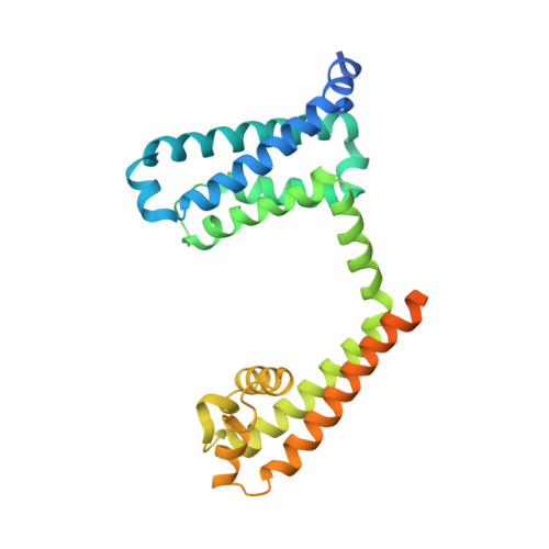
NavAb NormoPP mutant


Experimental Data Snapshot

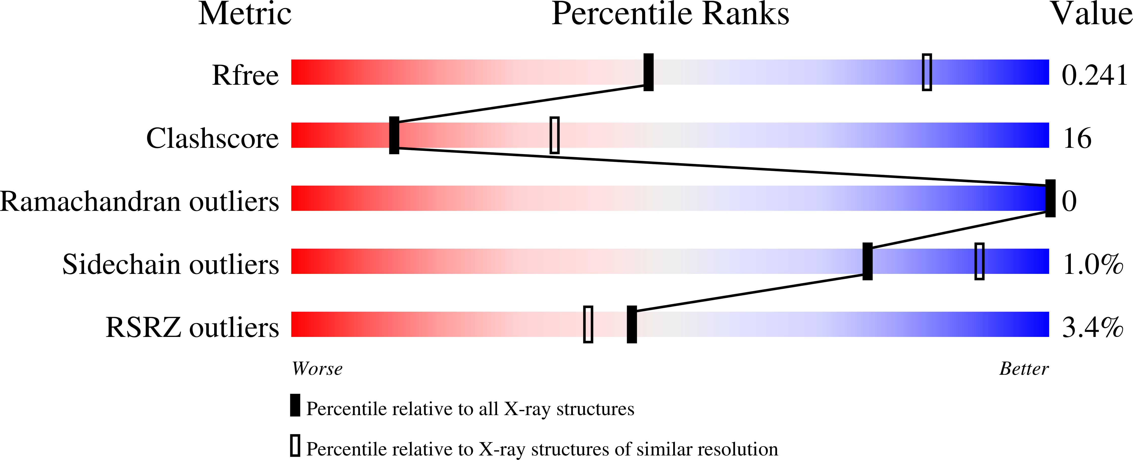
  • Method: X-RAY DIFFRACTION
  • Resolution: 2.86 Å
  • R-Value Free: 0.240 
  • R-Value Work: 0.212 
  • R-Value Observed: 0.214 

Starting Model: experimental
View more details

wwPDB Validation   3D Report Full Report


Ligand Structure Quality Assessment 


This is version 1.4 of the entry. See complete history


Literature

Structural basis for gating pore current in periodic paralysis.

Jiang, D.Gamal El-Din, T.M.Ing, C.Lu, P.Pomes, R.Zheng, N.Catterall, W.A.

(2018) Nature 557: 590-594

  • DOI: https://doi.org/10.1038/s41586-018-0120-4
  • Primary Citation of Related Structures:  
    6C1E, 6C1K, 6C1M, 6C1P

  • PubMed Abstract: 

    Potassium-sensitive hypokalaemic and normokalaemic periodic paralysis are inherited skeletal muscle diseases characterized by episodes of flaccid muscle weakness 1,2 . They are caused by single mutations in positively charged residues ('gating charges') in the S4 transmembrane segment of the voltage sensor of the voltage-gated sodium channel Na v 1.4 or the calcium channel Ca v 1.1 1,2 . Mutations of the outermost gating charges (R1 and R2) cause hypokalaemic periodic paralysis 1,2 by creating a pathogenic gating pore in the voltage sensor through which cations leak in the resting state 3,4 . Mutations of the third gating charge (R3) cause normokalaemic periodic paralysis 5 owing to cation leak in both activated and inactivated states 6 . Here we present high-resolution structures of the model bacterial sodium channel Na v Ab with the analogous gating-charge mutations 7,8 , which have similar functional effects as in the human channels. The R2G and R3G mutations have no effect on the backbone structures of the voltage sensor, but they create an aqueous cavity near the hydrophobic constriction site that controls gating charge movement through the voltage sensor. The R3G mutation extends the extracellular aqueous cleft through the entire length of the activated voltage sensor, creating an aqueous path through the membrane. Conversely, molecular modelling shows that the R2G mutation creates a continuous aqueous path through the membrane only in the resting state. Crystal structures of Na v Ab(R2G) in complex with guanidinium define a potential drug target site. Molecular dynamics simulations illustrate the mechanism of Na + permeation through the mutant gating pore in concert with conformational fluctuations of the gating charge R4. Our results reveal pathogenic mechanisms of periodic paralysis at the atomic level and suggest designs of drugs that may prevent ionic leak and provide symptomatic relief from hypokalaemic and normokalaemic periodic paralysis.


  • Organizational Affiliation

    Department of Pharmacology, University of Washington, Seattle, WA, USA.


Macromolecules
Find similar proteins by:  (by identity cutoff)  |  3D Structure
Entity ID: 1
MoleculeChains Sequence LengthOrganismDetailsImage
Ion transport protein
A, B
285Aliarcobacter butzleri RM4018Mutation(s): 2 
Gene Names: Abu_1752
Membrane Entity: Yes 
UniProt
Find proteins for A8EVM5 (Aliarcobacter butzleri (strain RM4018))
Explore A8EVM5 
Go to UniProtKB:  A8EVM5
Entity Groups  
Sequence Clusters30% Identity50% Identity70% Identity90% Identity95% Identity100% Identity
UniProt GroupA8EVM5
Sequence Annotations
Expand
  • Reference Sequence
Small Molecules
Ligands 3 Unique
IDChains Name / Formula / InChI Key2D Diagram3D Interactions
PX4
Query on PX4

Download Ideal Coordinates CCD File 
C [auth A]
D [auth A]
E [auth A]
F [auth A]
G [auth A]
C [auth A],
D [auth A],
E [auth A],
F [auth A],
G [auth A],
H [auth A],
I [auth A],
K [auth A],
L [auth A],
M [auth A],
N [auth B],
O [auth B],
P [auth B],
Q [auth B],
R [auth B],
S [auth B],
T [auth B],
U [auth B]
1,2-DIMYRISTOYL-SN-GLYCERO-3-PHOSPHOCHOLINE
C36 H73 N O8 P
CITHEXJVPOWHKC-UUWRZZSWSA-O
UHH
Query on UHH

Download Ideal Coordinates CCD File 
V [auth B](3ALPHA,5ALPHA,7ALPHA,8ALPHA,12ALPHA,14BETA,17ALPHA)-3,7,12-TRIHYDROXYCHOL-1-EN-24-AMIDE
C24 H39 N O4
FMNKZHQYXMWDCG-OELDTZBJSA-N
BNC
Query on BNC

Download Ideal Coordinates CCD File 
J [auth A]5-BETA-24-NOR-CHOLANE-3(ALPHA),7(ALPHA),12(ALPHA)-TRIOL
C23 H40 O3
HOGZQSSVBGJUMW-ZTSBSJDLSA-N
Experimental Data & Validation

Experimental Data

  • Method: X-RAY DIFFRACTION
  • Resolution: 2.86 Å
  • R-Value Free: 0.240 
  • R-Value Work: 0.212 
  • R-Value Observed: 0.214 
  • Space Group: I 2 2 2
Unit Cell:
Length ( Å )Angle ( ˚ )
a = 126.775α = 90
b = 126.997β = 90
c = 192.287γ = 90
Software Package:
Software NamePurpose
PHENIXrefinement
HKL-2000data reduction
HKL-2000data scaling
AutoSolphasing

Structure Validation

View Full Validation Report


Ligand Structure Quality Assessment 


Entry History & Funding Information

Deposition Data


Funding OrganizationLocationGrant Number
National Institutes of Health/National Human Genome Research Institute (NIH/NHGRI)United StatesNS015751

Revision History  (Full details and data files)

  • Version 1.0: 2018-05-16
    Type: Initial release
  • Version 1.1: 2018-05-30
    Changes: Data collection, Database references
  • Version 1.2: 2018-06-06
    Changes: Data collection, Database references
  • Version 1.3: 2019-12-18
    Changes: Author supporting evidence
  • Version 1.4: 2023-10-04
    Changes: Data collection, Database references, Refinement description