6E43

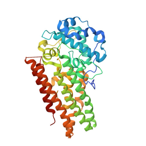
Crystal structure of human indoleamine 2,3-dioxygenase 1 (IDO1) in complex with a BMS-978587 analog


Experimental Data Snapshot

  • Method: X-RAY DIFFRACTION
  • Resolution: 1.71 Å
  • R-Value Free: 0.231 
  • R-Value Work: 0.194 
  • R-Value Observed: 0.194 

wwPDB Validation   3D Report Full Report


Ligand Structure Quality Assessment 


This is version 1.1 of the entry. See complete history


Literature

High-resolution structures of inhibitor complexes of human indoleamine 2,3-dioxygenase 1 in a new crystal form.

Luo, S.Xu, K.Xiang, S.Chen, J.Chen, C.Guo, C.Tong, Y.Tong, L.

(2018) Acta Crystallogr F Struct Biol Commun 74: 717-724

  • DOI: https://doi.org/10.1107/S2053230X18012955
  • Primary Citation of Related Structures:  
    6E40, 6E41, 6E42, 6E43, 6E44, 6E45, 6E46

  • PubMed Abstract: 

    Human indoleamine 2,3-dioxygenase 1 (IDO1) is a heme-dependent enzyme with important roles in many cellular processes and is a potential target for drug discovery against cancer and other diseases. Crystal structures of IDO1 in complex with various inhibitors have been reported. Many of these crystals belong to the same crystal form and most of the reported structures have resolutions in the range 3.2-2.3 Å. Here, three new crystal forms of human IDO1 obtained by introducing a surface mutation, K116A/K117A, distant from the active site are reported. One of these crystal forms diffracted to 1.5 Å resolution and can be readily used for soaking experiments to determine high-resolution structures of IDO1 in complex with the substrate tryptophan or inhibitors that coordinate the heme. In addition, this mutant was used to produce crystals of a complex with an inhibitor that targets the apo form of the enzyme under the same conditions; the structure of this complex was determined at 1.7 Å resolution. Overall, this mutant represents a robust platform for determining the structures of inhibitor and substrate complexes of IDO1 at high resolution.


  • Organizational Affiliation

    Department of Biological Sciences, Columbia University, New York, NY 10027, USA.


Macromolecules
Find similar proteins by:  (by identity cutoff)  |  3D Structure
Entity ID: 1
MoleculeChains Sequence LengthOrganismDetailsImage
Indoleamine 2,3-dioxygenase 1
A, B, C, D
405Homo sapiensMutation(s): 2 
Gene Names: IDO1IDOINDO
EC: 1.13.11.52
UniProt & NIH Common Fund Data Resources
Find proteins for P14902 (Homo sapiens)
Explore P14902 
Go to UniProtKB:  P14902
PHAROS:  P14902
GTEx:  ENSG00000131203 
Entity Groups  
Sequence Clusters30% Identity50% Identity70% Identity90% Identity95% Identity100% Identity
UniProt GroupP14902
Sequence Annotations
Expand
  • Reference Sequence
Small Molecules
Ligands 2 Unique
IDChains Name / Formula / InChI Key2D Diagram3D Interactions
HQM
Query on HQM

Download Ideal Coordinates CCD File 
E [auth A],
G [auth B],
I [auth C],
K [auth D]
(1R,2S)-2-(4-[cyclohexyl(2-methylpropyl)amino]-3-{[(4-methylphenyl)carbamoyl]amino}phenyl)cyclopropane-1-carboxylic acid
C28 H37 N3 O3
AHNMZQOTEVUTJH-DNQXCXABSA-N
BEZ
Query on BEZ

Download Ideal Coordinates CCD File 
F [auth A],
H [auth B],
J [auth C],
L [auth D]
BENZOIC ACID
C7 H6 O2
WPYMKLBDIGXBTP-UHFFFAOYSA-N
Experimental Data & Validation

Experimental Data

  • Method: X-RAY DIFFRACTION
  • Resolution: 1.71 Å
  • R-Value Free: 0.231 
  • R-Value Work: 0.194 
  • R-Value Observed: 0.194 
  • Space Group: P 21 21 2
Unit Cell:
Length ( Å )Angle ( ˚ )
a = 80.481α = 90
b = 195.388β = 90
c = 118.223γ = 90
Software Package:
Software NamePurpose
PHENIXrefinement
XSCALEdata scaling
PDB_EXTRACTdata extraction
XDSdata reduction
PHENIXphasing

Structure Validation

View Full Validation Report


Ligand Structure Quality Assessment 


Entry History 

Deposition Data

Revision History  (Full details and data files)

  • Version 1.0: 2018-11-14
    Type: Initial release
  • Version 1.1: 2024-03-13
    Changes: Data collection, Database references