Altered structure and dynamics of pathogenic cytochrome c variants correlate with increased apoptotic activity.
Fellner, M., Parakra, R., McDonald, K.O., Kass, I., Jameson, G.N.L., Wilbanks, S.M., Ledgerwood, E.C.(2021) Biochem J
- PubMed: 33480393
- DOI: https://doi.org/10.1042/BCJ20200793
- Primary Citation of Related Structures:
5EXQ, 6ECJ - PubMed Abstract:
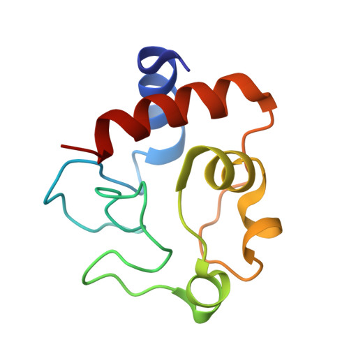
Mutation of cytochrome c in humans causes mild autosomal dominant thrombocytopenia. The role of cytochrome c in platelet formation, and the molecular mechanism underlying the association of cytochrome c mutations with thrombocytopenia remains unknown, although a gain-of-function is most likely. Cytochrome c contributes to several cellular processes, with an exchange between conformational states proposed to regulate changes in function. Here, we use experimental and computational approaches to determine whether pathogenic variants share changes in structure and function, and to understand how these changes might occur. Three pathogenic variants (G41S, Y48H, A51V) cause an increase in apoptosome activation and peroxidase activity. Molecular dynamics simulations of these variants, and two non-naturally occurring variants (G41A, G41T), indicate that increased apoptosome activation correlates with the increased overall flexibility of cytochrome c, particularly movement of the Ω loops. Crystal structures of Y48H and G41T complement these studies which overall suggest that the binding of cytochrome c to apoptotic protease activating factor-1 (Apaf-1) may involve an 'induced fit' mechanism which is enhanced in the more conformationally mobile variants. In contrast, peroxidase activity did not significantly correlate with protein dynamics. Thus, the mechanism by which the variants increase peroxidase activity is not related to the conformational dynamics of the native hexacoordinate state of cytochrome c. Recent molecular dynamics data proposing conformational mobility of specific cytochrome c regions underpins changes in reduction potential and alkaline transition pK was not fully supported. These data highlight that conformational dynamics of cytochrome c drive some but not all of its properties and activities.
Organizational Affiliation:
Department of Biochemistry, School of Biomedical Sciences, University of Otago, Dunedin, New Zealand.


