6EU5

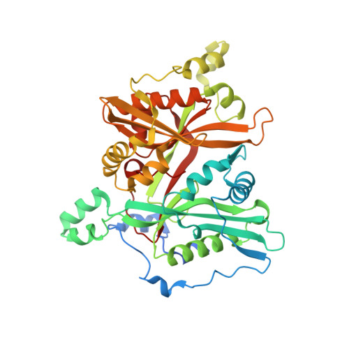
Leishmania major N-myristoyltransferase with bound myristoyl-CoA and inhibitor


Experimental Data Snapshot

  • Method: X-RAY DIFFRACTION
  • Resolution: 1.50 Å
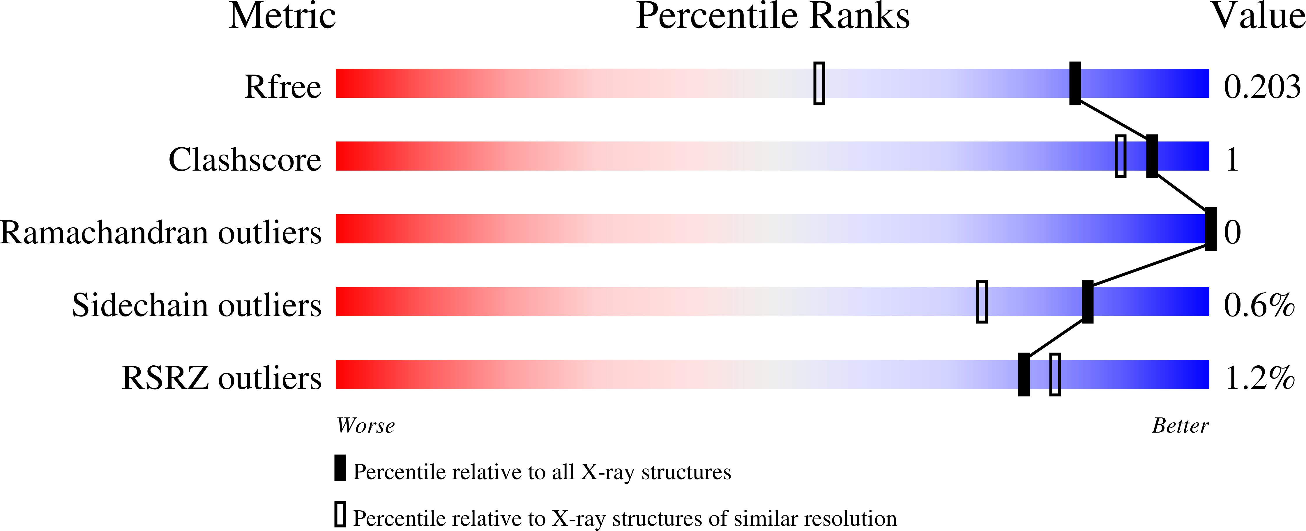
  • R-Value Free: 0.203 
  • R-Value Work: 0.169 
  • R-Value Observed: 0.171 

Starting Model: experimental
View more details

wwPDB Validation   3D Report Full Report


Ligand Structure Quality Assessment 


This is version 1.2 of the entry. See complete history


Literature

How To Design Selective Ligands for Highly Conserved Binding Sites: A Case Study UsingN-Myristoyltransferases as a Model System.

Kersten, C.Fleischer, E.Kehrein, J.Borek, C.Jaenicke, E.Sotriffer, C.Brenk, R.

(2019) J Med Chem 

  • DOI: https://doi.org/10.1021/acs.jmedchem.9b00586
  • Primary Citation of Related Structures:  
    6EU5, 6EWF, 6F56, 6FZ2, 6FZ3, 6FZ5

  • PubMed Abstract: 

    A model system of two related enzymes with conserved binding sites, namely N -myristoyltransferase from two different organisms, was studied to decipher the driving forces that lead to selective inhibition in such cases. Using a combination of computational and experimental tools, two different selectivity-determining features were identified. For some ligands, a change in side-chain flexibility appears to be responsible for selective inhibition. Remarkably, this was observed for residues orienting their side chains away from the ligands. For other ligands, selectivity is caused by interfering with a water molecule that binds more strongly to the off-target than to the target. On the basis of this finding, a virtual screen for selective compounds was conducted, resulting in three hit compounds with the desired selectivity profile. This study delivers a guideline on how to assess selectivity-determining features in proteins with conserved binding sites and to translate this knowledge into the design of selective inhibitors.


  • Organizational Affiliation

    Institute of Pharmacy and Biochemistry, Johannes Gutenberg-Universität Mainz, Staudingerweg 5, 55128 Mainz, Germany.


Macromolecules
Find similar proteins by:  (by identity cutoff)  |  3D Structure
Entity ID: 1
MoleculeChains Sequence LengthOrganismDetailsImage
Glycylpeptide N-tetradecanoyltransferase438Leishmania majorMutation(s): 0 
Gene Names: NMTLMJF_32_0080
EC: 2.3.1.97
UniProt
Find proteins for Q4Q5S8 (Leishmania major)
Explore Q4Q5S8 
Go to UniProtKB:  Q4Q5S8
Entity Groups  
Sequence Clusters30% Identity50% Identity70% Identity90% Identity95% Identity100% Identity
UniProt GroupQ4Q5S8
Sequence Annotations
Expand
  • Reference Sequence
Small Molecules
Ligands 2 Unique
IDChains Name / Formula / InChI Key2D Diagram3D Interactions
MYA
Query on MYA

Download Ideal Coordinates CCD File 
B [auth A]TETRADECANOYL-COA
C35 H62 N7 O17 P3 S
DUAFKXOFBZQTQE-QSGBVPJFSA-N
BXN (Subject of Investigation/LOI)
Query on BXN

Download Ideal Coordinates CCD File 
C [auth A]4-[3-[(8~{a}~{R})-3,4,6,7,8,8~{a}-hexahydro-1~{H}-pyrrolo[1,2-a]pyrazin-2-yl]propyl]-2,6-bis(chloranyl)-~{N}-methyl-~{N}-(1,3,5-trimethylpyrazol-4-yl)benzenesulfonamide
C23 H33 Cl2 N5 O2 S
IMQPHOQRHBIWQQ-LJQANCHMSA-N
Experimental Data & Validation

Experimental Data

  • Method: X-RAY DIFFRACTION
  • Resolution: 1.50 Å
  • R-Value Free: 0.203 
  • R-Value Work: 0.169 
  • R-Value Observed: 0.171 
  • Space Group: P 1 21 1
Unit Cell:
Length ( Å )Angle ( ˚ )
a = 48.77α = 90
b = 90.59β = 114.141
c = 53.42γ = 90
Software Package:
Software NamePurpose
autoPROCdata processing
PHASERphasing
Cootmodel building
PHENIXrefinement
XDSdata reduction
XSCALEdata scaling

Structure Validation

View Full Validation Report


Ligand Structure Quality Assessment 


Entry History & Funding Information

Deposition Data


Funding OrganizationLocationGrant Number
German Research FoundationGermany--

Revision History  (Full details and data files)

  • Version 1.0: 2019-02-06
    Type: Initial release
  • Version 1.1: 2019-08-28
    Changes: Data collection, Database references
  • Version 1.2: 2024-01-17
    Changes: Advisory, Data collection, Database references, Refinement description