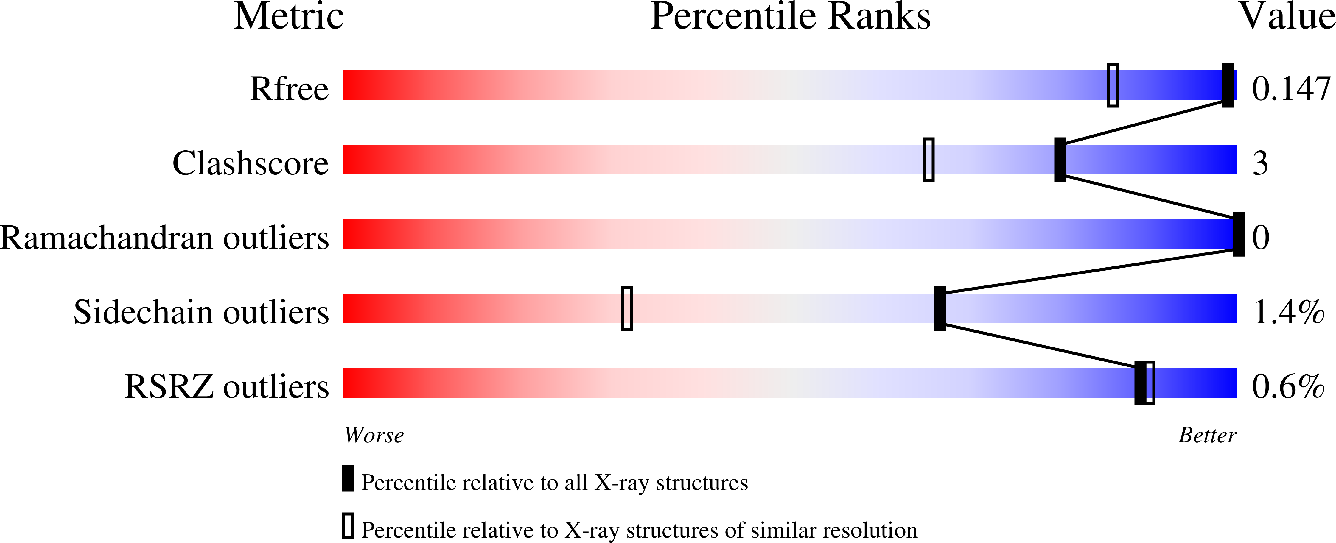
6FEA

A. vinelandii vanadium nitrogenase, turnover state


Experimental Data Snapshot

  • Method: X-RAY DIFFRACTION
  • Resolution: 1.20 Å
  • R-Value Free: 0.145 
  • R-Value Work: 0.117 
  • R-Value Observed: 0.118 

Starting Model: experimental
View more details

wwPDB Validation   3D Report Full Report


Ligand Structure Quality Assessment 


This is version 1.2 of the entry. See complete history


Literature

A bound reaction intermediate sheds light on the mechanism of nitrogenase.

Sippel, D.Rohde, M.Netzer, J.Trncik, C.Gies, J.Grunau, K.Djurdjevic, I.Decamps, L.Andrade, S.L.A.Einsle, O.

(2018) Science 359: 1484-1489

  • DOI: https://doi.org/10.1126/science.aar2765
  • Primary Citation of Related Structures:  
    6FEA

  • PubMed Abstract: 

    Reduction of N 2 by nitrogenases occurs at an organometallic iron cofactor that commonly also contains either molybdenum or vanadium. The well-characterized resting state of the cofactor does not bind substrate, so its mode of action remains enigmatic. Carbon monoxide was recently found to replace a bridging sulfide, but the mechanistic relevance was unclear. Here we report the structural analysis of vanadium nitrogenase with a bound intermediate, interpreted as a μ 2 -bridging, protonated nitrogen that implies the site and mode of substrate binding to the cofactor. Binding results in a flip of amino acid glutamine 176, which hydrogen-bonds the ligand and creates a holding position for the displaced sulfide. The intermediate likely represents state E 6 or E 7 of the Thorneley-Lowe model and provides clues to the remainder of the catalytic cycle.


  • Organizational Affiliation

    Institut für Biochemie, Albert-Ludwigs-Universität Freiburg, Albertstraße 21, 79104 Freiburg, Germany.


Macromolecules
Find similar proteins by:  (by identity cutoff)  |  3D Structure
Entity ID: 1
MoleculeChains Sequence LengthOrganismDetailsImage
Nitrogenase protein alpha chain
A, D
474Azotobacter vinelandii DJMutation(s): 0 
EC: 1.18.6.1
UniProt
Find proteins for C1DI25 (Azotobacter vinelandii (strain DJ / ATCC BAA-1303))
Explore C1DI25 
Go to UniProtKB:  C1DI25
Entity Groups  
Sequence Clusters30% Identity50% Identity70% Identity90% Identity95% Identity100% Identity
UniProt GroupC1DI25
Sequence Annotations
Expand
  • Reference Sequence
Find similar proteins by:  (by identity cutoff)  |  3D Structure
Entity ID: 2
MoleculeChains Sequence LengthOrganismDetailsImage
Vanadium nitrogenase beta subunit, vnfK
B, E
475Azotobacter vinelandii DJMutation(s): 0 
EC: 1.18.6.1
UniProt
Find proteins for C1DI23 (Azotobacter vinelandii (strain DJ / ATCC BAA-1303))
Explore C1DI23 
Go to UniProtKB:  C1DI23
Entity Groups  
Sequence Clusters30% Identity50% Identity70% Identity90% Identity95% Identity100% Identity
UniProt GroupC1DI23
Sequence Annotations
Expand
  • Reference Sequence
Find similar proteins by:  (by identity cutoff)  |  3D Structure
Entity ID: 3
MoleculeChains Sequence LengthOrganismDetailsImage
Vanadium nitrogenase, delta subunit, VnfG
C, F
113Azotobacter vinelandii DJMutation(s): 0 
EC: 1.18.6.1
UniProt
Find proteins for C1DI24 (Azotobacter vinelandii (strain DJ / ATCC BAA-1303))
Explore C1DI24 
Go to UniProtKB:  C1DI24
Entity Groups  
Sequence Clusters30% Identity50% Identity70% Identity90% Identity95% Identity100% Identity
UniProt GroupC1DI24
Sequence Annotations
Expand
  • Reference Sequence
Small Molecules
Ligands 7 Unique
IDChains Name / Formula / InChI Key2D Diagram3D Interactions
D6N
Query on D6N

Download Ideal Coordinates CCD File 
H [auth A],
Q [auth D]
FeV
C Fe7 N S7 V
YNVNIECPQFRAMK-UHFFFAOYSA-N
CLF
Query on CLF

Download Ideal Coordinates CCD File 
L [auth A],
U [auth D]
FE(8)-S(7) CLUSTER
Fe8 S7
JKVMXLBGZBULKV-UHFFFAOYSA-N
HCA
Query on HCA

Download Ideal Coordinates CCD File 
G [auth A],
R [auth D]
3-HYDROXY-3-CARBOXY-ADIPIC ACID
C7 H10 O7
XKJVEVRQMLKSMO-SSDOTTSWSA-N
ZN
Query on ZN

Download Ideal Coordinates CCD File 
K [auth A],
O [auth B]
ZINC ION
Zn
PTFCDOFLOPIGGS-UHFFFAOYSA-N
CO3
Query on CO3

Download Ideal Coordinates CCD File 
I [auth A],
S [auth D]
CARBONATE ION
C O3
BVKZGUZCCUSVTD-UHFFFAOYSA-L
H2S
Query on H2S

Download Ideal Coordinates CCD File 
J [auth A],
T [auth D]
HYDROSULFURIC ACID
H2 S
RWSOTUBLDIXVET-UHFFFAOYSA-N
MG
Query on MG

Download Ideal Coordinates CCD File 
M [auth B],
N [auth B],
P [auth C],
V [auth F]
MAGNESIUM ION
Mg
JLVVSXFLKOJNIY-UHFFFAOYSA-N
Experimental Data & Validation

Experimental Data

  • Method: X-RAY DIFFRACTION
  • Resolution: 1.20 Å
  • R-Value Free: 0.145 
  • R-Value Work: 0.117 
  • R-Value Observed: 0.118 
  • Space Group: P 1
Unit Cell:
Length ( Å )Angle ( ˚ )
a = 75.61α = 84.05
b = 79.75β = 72.44
c = 107.16γ = 75.25
Software Package:
Software NamePurpose
REFMACrefinement
XDSdata reduction
SCALAdata scaling
MOLREPphasing

Structure Validation

View Full Validation Report


Ligand Structure Quality Assessment 


Entry History & Funding Information

Deposition Data


Funding OrganizationLocationGrant Number
German Research FoundationGermanyRTG 1976
German Research FoundationGermanyPP 1927
European Research CouncilGermany310656

Revision History  (Full details and data files)

  • Version 1.0: 2018-03-28
    Type: Initial release
  • Version 1.1: 2018-04-11
    Changes: Data collection, Database references
  • Version 1.2: 2024-01-17
    Changes: Data collection, Database references, Derived calculations, Refinement description