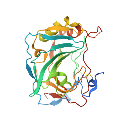
6G98

Three dimensional structure of human carbonic anhydrase IX in complex with sulfonamide


Experimental Data Snapshot

  • Method: X-RAY DIFFRACTION
  • Resolution: 2.47 Å
  • R-Value Free: 0.209 
  • R-Value Work: 0.169 
  • R-Value Observed: 0.171 

Starting Model: experimental
View more details

wwPDB Validation   3D Report Full Report


Ligand Structure Quality Assessment 


This is version 1.2 of the entry. See complete history


Literature

Novel fluorinated carbonic anhydrase IX inhibitors reduce hypoxia-induced acidification and clonogenic survival of cancer cells.

Kazokaite, J.Niemans, R.Dudutiene, V.Becker, H.M.Leitans, J.Zubriene, A.Baranauskiene, L.Gondi, G.Zeidler, R.Matuliene, J.Tars, K.Yaromina, A.Lambin, P.Dubois, L.J.Matulis, D.

(2018) Oncotarget 9: 26800-26816

  • DOI: https://doi.org/10.18632/oncotarget.25508
  • Primary Citation of Related Structures:  
    6FE0, 6FE1, 6FE2, 6G98, 6G9U

  • PubMed Abstract: 

    Human carbonic anhydrase (CA) IX has emerged as a promising anticancer target and a diagnostic biomarker for solid hypoxic tumors. Novel fluorinated CA IX inhibitors exhibited up to 50 pM affinity towards the recombinant human CA IX, selectivity over other CAs, and direct binding to Zn(II) in the active site of CA IX inducing novel conformational changes as determined by X-ray crystallography. Mass spectrometric gas-analysis confirmed the CA IX-based mechanism of the inhibitors in a CRISPR/Cas9-mediated CA IX knockout in HeLa cells. Hypoxia-induced extracellular acidification was significantly reduced in HeLa, H460, MDA-MB-231, and A549 cells exposed to the compounds, with the IC 50 values up to 1.29 nM. A decreased clonogenic survival was observed when hypoxic H460 3D spheroids were incubated with our lead compound. These novel compounds are therefore promising agents for CA IX-specific therapy.


  • Organizational Affiliation

    Department of Biothermodynamics and Drug Design, Institute of Biotechnology, Vilnius University, Vilnius, Lithuania.


Macromolecules
Find similar proteins by:  (by identity cutoff)  |  3D Structure
Entity ID: 1
MoleculeChains Sequence LengthOrganismDetailsImage
Carbonic anhydrase 9
A, B, C, D
257Homo sapiensMutation(s): 1 
Gene Names: CA9G250MN
EC: 4.2.1.1
UniProt & NIH Common Fund Data Resources
Find proteins for Q16790 (Homo sapiens)
Explore Q16790 
Go to UniProtKB:  Q16790
PHAROS:  Q16790
GTEx:  ENSG00000107159 
Entity Groups  
Sequence Clusters30% Identity50% Identity70% Identity90% Identity95% Identity100% Identity
UniProt GroupQ16790
Sequence Annotations
Expand
  • Reference Sequence
Small Molecules
Ligands 2 Unique
IDChains Name / Formula / InChI Key2D Diagram3D Interactions
ER5 (Subject of Investigation/LOI)
Query on ER5

Download Ideal Coordinates CCD File 
F [auth A],
H [auth B],
J [auth C],
L [auth D]
4-[2-[3-(cyclododecylamino)-2,5,6-tris(fluoranyl)-4-sulfamoyl-phenyl]sulfanylethyl]benzoic acid
C27 H35 F3 N2 O4 S2
BMKRNBDPIOXDOC-UHFFFAOYSA-N
ZN
Query on ZN

Download Ideal Coordinates CCD File 
E [auth A],
G [auth B],
I [auth C],
K [auth D]
ZINC ION
Zn
PTFCDOFLOPIGGS-UHFFFAOYSA-N
Experimental Data & Validation

Experimental Data

  • Method: X-RAY DIFFRACTION
  • Resolution: 2.47 Å
  • R-Value Free: 0.209 
  • R-Value Work: 0.169 
  • R-Value Observed: 0.171 
  • Space Group: H 3
Unit Cell:
Length ( Å )Angle ( ˚ )
a = 152.39α = 90
b = 152.39β = 90
c = 171.83γ = 120
Software Package:
Software NamePurpose
REFMACrefinement
SCALAdata scaling

Structure Validation

View Full Validation Report


Ligand Structure Quality Assessment 


Entry History 

Deposition Data

Revision History  (Full details and data files)

  • Version 1.0: 2018-07-04
    Type: Initial release
  • Version 1.1: 2024-01-17
    Changes: Data collection, Database references, Derived calculations, Refinement description
  • Version 1.2: 2024-10-16
    Changes: Structure summary