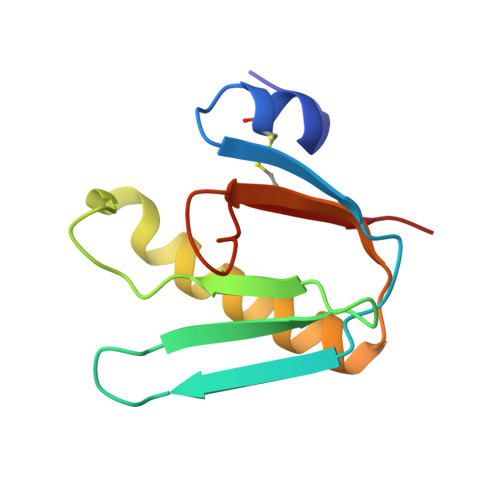
Crystal structures of cholera toxin in complex with fucosylated receptors point to importance of secondary binding site.
Heim, J.B., Hodnik, V., Heggelund, J.E., Anderluh, G., Krengel, U.(2019) Sci Rep 9: 12243-12243
- PubMed: 31439922
- DOI: https://doi.org/10.1038/s41598-019-48579-2
- Primary Citation of Related Structures:
6HJD, 6HMW, 6HMY - PubMed Abstract:
Cholera is a life-threatening diarrhoeal disease caused by the human pathogen Vibrio cholerae. Infection occurs after ingestion of the bacteria, which colonize the human small intestine and secrete their major virulence factor - the cholera toxin (CT). The GM1 ganglioside is considered the primary receptor of the CT, but recent studies suggest that also fucosylated receptors such as histo-blood group antigens are important for cellular uptake and toxicity. Recently, a special focus has been on the histo-blood group antigen Lewis x (Le x ), however, where and how the CT binds to Le x remains unclear. Here we report the high-resolution crystal structure (1.5 Å) of the receptor-binding B-subunits of the CT bound to the Le x trisaccharide, and complementary quantitative binding data for CT holotoxins. Le x , and also L-fucose alone, bind to the secondary binding site of the toxin, distinct from the GM1 binding site. In contrast, fucosyl-GM1 mainly binds to the primary binding site due to high-affinity interactions of its GM1 core. Le x is the first histo-blood group antigen of non-secretor phenotype structurally investigated in complex with CT. Together with the quantitative binding data, this allows unique insight into why individuals with non-secretor phenotype are more prone to severe cholera than so-called 'secretors'.
Organizational Affiliation:
Department of Chemistry, University of Oslo, P.O. Box 1033, NO-0315, Blindern, Norway.


