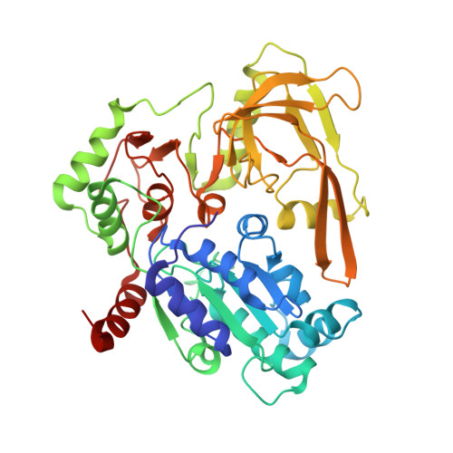
6HPU

Crystal structure of human Pif1 helicase in complex with ADP-AlF4


Experimental Data Snapshot

  • Method: X-RAY DIFFRACTION
  • Resolution: 3.96 Å
  • R-Value Free: 0.253 
  • R-Value Work: 0.175 
  • R-Value Observed: 0.179 

Starting Model: experimental
View more details

wwPDB Validation   3D Report Full Report


Ligand Structure Quality Assessment 


This is version 1.3 of the entry. See complete history


Literature

Structural and functional analysis of the nucleotide and DNA binding activities of the human PIF1 helicase.

Dehghani-Tafti, S.Levdikov, V.Antson, A.A.Bax, B.Sanders, C.M.

(2019) Nucleic Acids Res 47: 3208-3222

  • DOI: https://doi.org/10.1093/nar/gkz028
  • Primary Citation of Related Structures:  
    6HPH, 6HPQ, 6HPT, 6HPU

  • PubMed Abstract: 

    Pif1 is a multifunctional helicase and DNA processing enzyme that has roles in genome stability. The enzyme is conserved in eukaryotes and also found in some prokaryotes. The functions of human PIF1 (hPIF1) are also critical for survival of certain tumour cell lines during replication stress, making it an important target for cancer therapy. Crystal structures of hPIF1 presented here explore structural events along the chemical reaction coordinate of ATP hydrolysis at an unprecedented level of detail. The structures for the apo as well as the ground and transition states reveal conformational adjustments in defined protein segments that can trigger larger domain movements required for helicase action. Comparisons with the structures of yeast and bacterial Pif1 reveal a conserved ssDNA binding channel in hPIF1 that we show is critical for single-stranded DNA binding during unwinding, but not the binding of G quadruplex DNA. Mutational analysis suggests that while the ssDNA-binding channel is important for helicase activity, it is not used in DNA annealing. Structural differences, in particular in the DNA strand separation wedge region, highlight significant evolutionary divergence of the human PIF1 protein from bacterial and yeast orthologues.


  • Organizational Affiliation

    Department of Oncology and Metabolism, Academic Unit of Molecular Oncology, University of Sheffield, Beech Hill Rd., Sheffield S10 2RX, UK.


Macromolecules
Find similar proteins by:  (by identity cutoff)  |  3D Structure
Entity ID: 1
MoleculeChains Sequence LengthOrganismDetailsImage
ATP-dependent DNA helicase PIF1
A, B
418Homo sapiensMutation(s): 0 
Gene Names: PIF1C15orf20
EC: 3.6.4.12
UniProt & NIH Common Fund Data Resources
Find proteins for Q9H611 (Homo sapiens)
Explore Q9H611 
Go to UniProtKB:  Q9H611
PHAROS:  Q9H611
GTEx:  ENSG00000140451 
Entity Groups  
Sequence Clusters30% Identity50% Identity70% Identity90% Identity95% Identity100% Identity
UniProt GroupQ9H611
Sequence Annotations
Expand
  • Reference Sequence
Experimental Data & Validation

Experimental Data

  • Method: X-RAY DIFFRACTION
  • Resolution: 3.96 Å
  • R-Value Free: 0.253 
  • R-Value Work: 0.175 
  • R-Value Observed: 0.179 
  • Space Group: P 32 2 1
Unit Cell:
Length ( Å )Angle ( ˚ )
a = 209.85α = 90
b = 209.85β = 90
c = 78.885γ = 120
Software Package:
Software NamePurpose
REFMACrefinement
xia2data reduction
xia2data scaling
MOLREPphasing

Structure Validation

View Full Validation Report


Ligand Structure Quality Assessment 


Entry History 

Deposition Data

Revision History  (Full details and data files)

  • Version 1.0: 2019-01-23
    Type: Initial release
  • Version 1.1: 2019-02-13
    Changes: Data collection, Database references
  • Version 1.2: 2019-04-17
    Changes: Data collection, Database references
  • Version 1.3: 2024-01-24
    Changes: Data collection, Database references, Refinement description