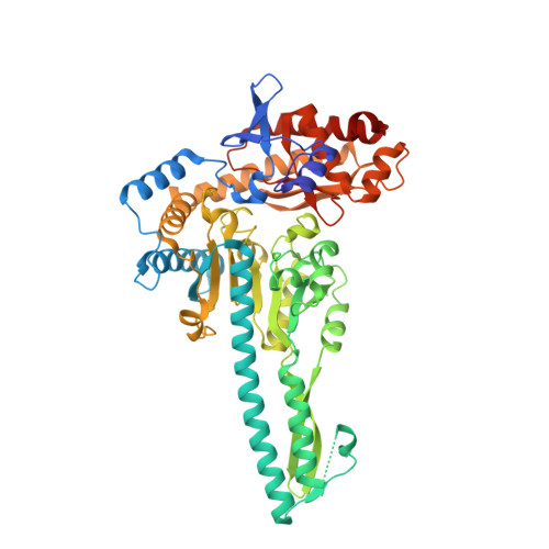
6JC9

Crystal structure of aminotransferase CrmG from Actinoalloteichus sp. WH1-2216-6 in complex with amino donor L-Gln


Experimental Data Snapshot

  • Method: X-RAY DIFFRACTION
  • Resolution: 2.35 Å
  • R-Value Free: 0.207 
  • R-Value Work: 0.172 
  • R-Value Observed: 0.173 

Starting Model: experimental
View more details

wwPDB Validation   3D Report Full Report


Ligand Structure Quality Assessment 


This is version 1.2 of the entry. See complete history


Literature

Structural studies reveal flexible roof of active site responsible for omega-transaminase CrmG overcoming by-product inhibition.

Xu, J.Tang, X.Zhu, Y.Yu, Z.Su, K.Zhang, Y.Dong, Y.Zhu, W.Zhang, C.Wu, R.Liu, J.

(2020) Commun Biol 3: 455-455

  • DOI: https://doi.org/10.1038/s42003-020-01184-w
  • Primary Citation of Related Structures:  
    6JC7, 6JC8, 6JC9, 6JCA, 6JCB

  • PubMed Abstract: 

    Amine compounds biosynthesis using ω-transaminases has received considerable attention in the pharmaceutical industry. However, the application of ω-transaminases was hampered by the fundamental challenge of severe by-product inhibition. Here, we report that ω-transaminase CrmG from Actinoalloteichus cyanogriseus WH1-2216-6 is insensitive to inhibition from by-product α-ketoglutarate or pyruvate. Combined with structural and QM/MM studies, we establish the detailed catalytic mechanism for CrmG. Our structural and biochemical studies reveal that the roof of the active site in PMP-bound CrmG is flexible, which will facilitate the PMP or by-product to dissociate from PMP-bound CrmG. Our results also show that amino acceptor caerulomycin M (CRM M), but not α-ketoglutarate or pyruvate, can form strong interactions with the roof of the active site in PMP-bound CrmG. Based on our results, we propose that the flexible roof of the active site in PMP-bound CrmG may facilitate CrmG to overcome inhibition from the by-product.


  • Organizational Affiliation

    State Key Laboratory of Respiratory Disease, Guangzhou Institutes of Biomedicine and Health, Chinese Academy of Sciences, 510530, Guangzhou, China. [email protected].


Macromolecules
Find similar proteins by:  (by identity cutoff)  |  3D Structure
Entity ID: 1
MoleculeChains Sequence LengthOrganismDetailsImage
CrmG
A, B, C, D
523Actinoalloteichus sp. WH1-2216-6Mutation(s): 0 
UniProt
Find proteins for H8Y6N2 (Actinoalloteichus sp. WH1-2216-6)
Explore H8Y6N2 
Go to UniProtKB:  H8Y6N2
Entity Groups  
Sequence Clusters30% Identity50% Identity70% Identity90% Identity95% Identity100% Identity
UniProt GroupH8Y6N2
Sequence Annotations
Expand
  • Reference Sequence
Small Molecules
Ligands 4 Unique
IDChains Name / Formula / InChI Key2D Diagram3D Interactions
PLP
Query on PLP

Download Ideal Coordinates CCD File 
E [auth A],
F [auth B],
H [auth C],
K [auth D]
PYRIDOXAL-5'-PHOSPHATE
C8 H10 N O6 P
NGVDGCNFYWLIFO-UHFFFAOYSA-N
GLN
Query on GLN

Download Ideal Coordinates CCD File 
J [auth C]GLUTAMINE
C5 H10 N2 O3
ZDXPYRJPNDTMRX-VKHMYHEASA-N
GOL
Query on GOL

Download Ideal Coordinates CCD File 
I [auth C]GLYCEROL
C3 H8 O3
PEDCQBHIVMGVHV-UHFFFAOYSA-N
ACY
Query on ACY

Download Ideal Coordinates CCD File 
G [auth B]ACETIC ACID
C2 H4 O2
QTBSBXVTEAMEQO-UHFFFAOYSA-N
Experimental Data & Validation

Experimental Data

  • Method: X-RAY DIFFRACTION
  • Resolution: 2.35 Å
  • R-Value Free: 0.207 
  • R-Value Work: 0.172 
  • R-Value Observed: 0.173 
  • Space Group: P 1
Unit Cell:
Length ( Å )Angle ( ˚ )
a = 84.039α = 106.48
b = 83.879β = 109.17
c = 88.41γ = 94.98
Software Package:
Software NamePurpose
Aimlessdata scaling
MOLREPphasing
REFMACrefinement
PDB_EXTRACTdata extraction
iMOSFLMdata reduction

Structure Validation

View Full Validation Report


Ligand Structure Quality Assessment 


Entry History & Funding Information

Deposition Data

  • Released Date: 2020-02-05 
  • Deposition Author(s): Xu, J., Liu, J.

Funding OrganizationLocationGrant Number
National Natural Science Foundation of ChinaChina31500638

Revision History  (Full details and data files)

  • Version 1.0: 2020-02-05
    Type: Initial release
  • Version 1.1: 2020-09-16
    Changes: Database references
  • Version 1.2: 2023-11-22
    Changes: Data collection, Database references, Refinement description