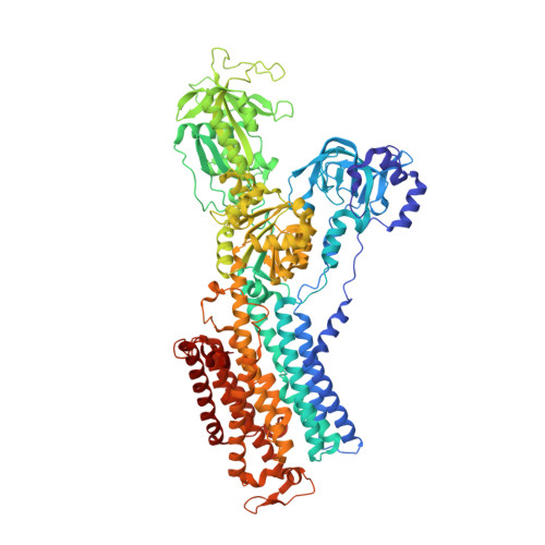
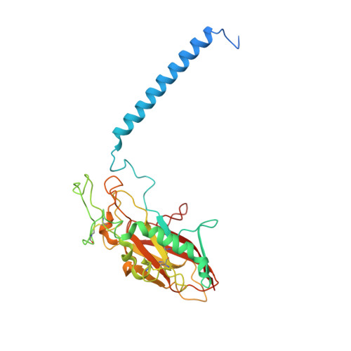
6JXJ

Rb+-bound E2-AlF state of the gastric proton pump (Tyr799Trp)


Experimental Data Snapshot

  • Method: X-RAY DIFFRACTION
  • Resolution: 2.50 Å
  • R-Value Free: 0.273 
  • R-Value Work: 0.214 
  • R-Value Observed: 0.217 

Starting Model: experimental
View more details

wwPDB Validation   3D Report Full Report


Ligand Structure Quality Assessment 


This is version 1.5 of the entry. See complete history


Literature

A single K + -binding site in the crystal structure of the gastric proton pump.

Yamamoto, K.Dubey, V.Irie, K.Nakanishi, H.Khandelia, H.Fujiyoshi, Y.Abe, K.

(2019) Elife 8

  • DOI: https://doi.org/10.7554/eLife.47701
  • Primary Citation of Related Structures:  
    6JXH, 6JXI, 6JXJ, 6JXK

  • PubMed Abstract: 

    The gastric proton pump (H + ,K + -ATPase), a P-type ATPase responsible for gastric acidification, mediates electro-neutral exchange of H + and K + coupled with ATP hydrolysis, but with an as yet undetermined transport stoichiometry. Here we show crystal structures at a resolution of 2.5 Å of the pump in the E2-P transition state, in which the counter-transporting cation is occluded. We found a single K + bound to the cation-binding site of the H + ,K + -ATPase, indicating an exchange of 1H + /1K + per hydrolysis of one ATP molecule. This fulfills the energy requirement for the generation of a six pH unit gradient across the membrane. The structural basis of K + recognition is resolved and supported by molecular dynamics simulations, establishing how the H + ,K + -ATPase overcomes the energetic challenge to generate an H + gradient of more than a million-fold-one of the highest cation gradients known in mammalian tissue-across the membrane.


  • Organizational Affiliation

    Cellular and Structural Physiology Institute, Nagoya University, Nagoya, Japan.


Macromolecules
Find similar proteins by:  (by identity cutoff)  |  3D Structure
Entity ID: 1
MoleculeChains Sequence LengthOrganismDetailsImage
Potassium-transporting ATPase alpha chain 1987Sus scrofaMutation(s): 4 
Gene Names: ATP4A
EC: 7.2.2.19
Membrane Entity: Yes 
UniProt
Find proteins for P19156 (Sus scrofa)
Explore P19156 
Go to UniProtKB:  P19156
Entity Groups  
Sequence Clusters30% Identity50% Identity70% Identity90% Identity95% Identity100% Identity
UniProt GroupP19156
Sequence Annotations
Expand
  • Reference Sequence
Find similar proteins by:  (by identity cutoff)  |  3D Structure
Entity ID: 2
MoleculeChains Sequence LengthOrganismDetailsImage
Potassium-transporting ATPase subunit beta289Sus scrofaMutation(s): 0 
Gene Names: ATP4B
Membrane Entity: Yes 
UniProt
Find proteins for P18434 (Sus scrofa)
Explore P18434 
Go to UniProtKB:  P18434
Entity Groups  
Sequence Clusters30% Identity50% Identity70% Identity90% Identity95% Identity100% Identity
UniProt GroupP18434
Glycosylation
Glycosylation Sites: 5
Sequence Annotations
Expand
  • Reference Sequence
Small Molecules
Ligands 7 Unique
IDChains Name / Formula / InChI Key2D Diagram3D Interactions
PCW
Query on PCW

Download Ideal Coordinates CCD File 
D [auth A],
E [auth A],
F [auth A],
G [auth A]
1,2-DIOLEOYL-SN-GLYCERO-3-PHOSPHOCHOLINE
C44 H85 N O8 P
SNKAWJBJQDLSFF-NVKMUCNASA-O
CE1
Query on CE1

Download Ideal Coordinates CCD File 
H [auth A]O-DODECANYL OCTAETHYLENE GLYCOL
C28 H58 O9
YYELLDKEOUKVIQ-UHFFFAOYSA-N
CLR
Query on CLR

Download Ideal Coordinates CCD File 
Q [auth B]CHOLESTEROL
C27 H46 O
HVYWMOMLDIMFJA-DPAQBDIFSA-N
NAG
Query on NAG

Download Ideal Coordinates CCD File 
R [auth B]
S [auth B]
T [auth B]
U [auth B]
V [auth B]
R [auth B],
S [auth B],
T [auth B],
U [auth B],
V [auth B],
W [auth B]
2-acetamido-2-deoxy-beta-D-glucopyranose
C8 H15 N O6
OVRNDRQMDRJTHS-FMDGEEDCSA-N
ALF
Query on ALF

Download Ideal Coordinates CCD File 
I [auth A]TETRAFLUOROALUMINATE ION
Al F4
UYOMQIYKOOHAMK-UHFFFAOYSA-J
RB
Query on RB

Download Ideal Coordinates CCD File 
J [auth A]
K [auth A]
L [auth A]
M [auth A]
N [auth A]
J [auth A],
K [auth A],
L [auth A],
M [auth A],
N [auth A],
O [auth A],
P [auth A],
X [auth B]
RUBIDIUM ION
Rb
NCCSSGKUIKYAJD-UHFFFAOYSA-N
MG
Query on MG

Download Ideal Coordinates CCD File 
C [auth A]MAGNESIUM ION
Mg
JLVVSXFLKOJNIY-UHFFFAOYSA-N
Experimental Data & Validation

Experimental Data

  • Method: X-RAY DIFFRACTION
  • Resolution: 2.50 Å
  • R-Value Free: 0.273 
  • R-Value Work: 0.214 
  • R-Value Observed: 0.217 
  • Space Group: P 31 2 1
Unit Cell:
Length ( Å )Angle ( ˚ )
a = 103.23α = 90
b = 103.23β = 90
c = 369.44γ = 120
Software Package:
Software NamePurpose
PHENIXrefinement
PHENIXrefinement
XDSdata reduction
XDSdata scaling
PHASERphasing

Structure Validation

View Full Validation Report


Ligand Structure Quality Assessment 


Entry History 

Deposition Data

Revision History  (Full details and data files)

  • Version 1.0: 2019-08-14
    Type: Initial release
  • Version 1.1: 2019-09-04
    Changes: Data collection, Database references
  • Version 1.2: 2020-07-29
    Type: Remediation
    Reason: Carbohydrate remediation
    Changes: Data collection, Derived calculations, Structure summary
  • Version 1.3: 2020-09-30
    Changes: Database references, Structure summary
  • Version 1.4: 2023-11-22
    Changes: Data collection, Database references, Refinement description
  • Version 1.5: 2024-11-13
    Changes: Structure summary