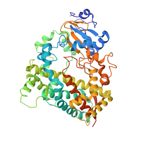
Interaction of Human Drug-Metabolizing CYP3A4 with Small Inhibitory Molecules.
Sevrioukova, I.(2019) Biochemistry 58: 930-939
- PubMed: 30676743
- DOI: https://doi.org/10.1021/acs.biochem.8b01221
- Primary Citation of Related Structures:
6MA6, 6MA7, 6MA8 - PubMed Abstract:
Binding of small inhibitory compounds to human cytochrome P450 3A4 (CYP3A4) could interfere with drug metabolism and lead to drug-drug interactions, the underlying mechanism of which is not fully understood due to insufficient structural information. This study investigated the interaction of recombinant CYP3A4 with a nonspecific inhibitor metyrapone, antifungal drug fluconazole, and protease inhibitor phenylmethanesulfonyl fluoride (PMSF). Metyrapone and fluconazole are classic type II ligands that inhibit CYP3A4 with medium strength by ligating to the heme iron, whereas PMSF, lacking the heme-ligating moiety, acts as a weak type I ligand and inhibitor of CYP3A4. High-resolution crystal structures revealed that the orientation of metyrapone is similar but not identical to that in the previously reported 1W0G model, whereas the flexible fluconazole adapts a conformer markedly different from that observed in the target CYP51 enzymes, which could explain its high potential for cross-reactivity. Besides hydrophobic and aromatic interactions with the heme and active site residues, both drugs establish water-mediated contacts that stabilize the inhibitory complexes. PMSF also binds near the catalytic center, with the phenyl group parallel to the heme. However, it does not displace the water ligand and is held in place via strong H-bonds formed by the sulfofluoride moiety with Ser119 and Arg212. Collectively, our data suggest that PMSF might have multiple binding sites and likely occupies the high-affinity site in the crystal structure. Moreover, its hydrolysis product, phenylmethanesulfonic acid, can also access and be retained in the CYP3A4 active site. Therefore, to avoid experimental artifacts, PMSF should be excluded from purification and assay solutions.
Organizational Affiliation:
Department of Molecular Biology and Biochemistry , University of California , Irvine , California 92697-3900 , United States.


