6NX9

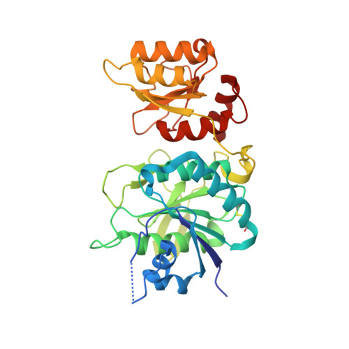
ECAII(D90T,K162T) MUTANT IN COMPLEX WITH CITRATE AT PH 7


Experimental Data Snapshot

  • Method: X-RAY DIFFRACTION
  • Resolution: 1.97 Å
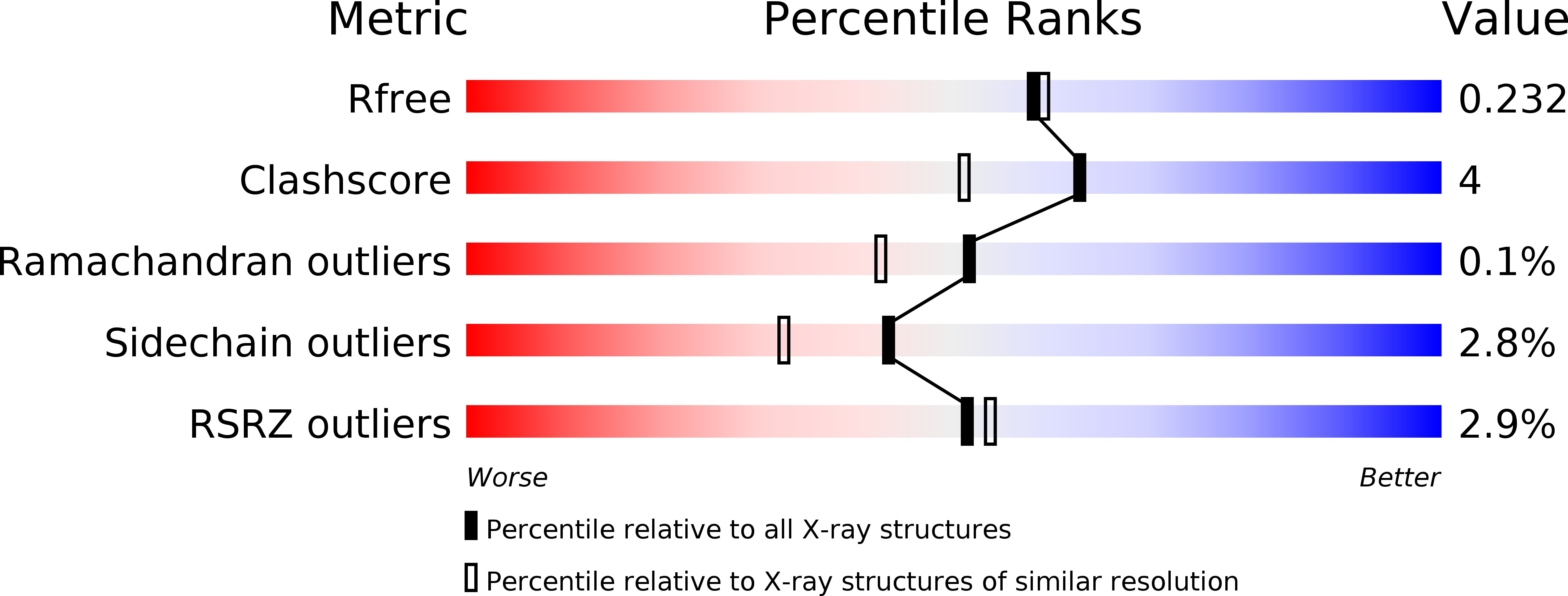
  • R-Value Free: 0.226 
  • R-Value Work: 0.160 
  • R-Value Observed: 0.162 

wwPDB Validation   3D Report Full Report


Ligand Structure Quality Assessment 


This is version 1.3 of the entry. See complete history


Literature

Opportunistic complexes of E. coli L-asparaginases with citrate anions.

Lubkowski, J.Chan, W.Wlodawer, A.

(2019) Sci Rep 9: 11070-11070

  • DOI: https://doi.org/10.1038/s41598-019-46432-0
  • Primary Citation of Related Structures:  
    6NX6, 6NX7, 6NX8, 6NX9, 6NXA, 6NXB, 6NXC, 6NXD

  • PubMed Abstract: 

    Active sites of enzymes are highly optimized for interactions with specific substrates, thus binding of opportunistic ligands is usually observed only in the absence of native substrates or products. However, during growth of crystals required for structure determination enzymes are often exposed to conditions significantly divergent from the native ones, leading to binding of unexpected ligands to active sites even in the presence of substrates. Failing to recognize this possibility may lead to incorrect interpretation of experimental results and to faulty conclusions. Here, we present several examples of binding of a citrate anion to the active sites of E. coli L-asparaginases I and II, even in the presence of the native substrate, L-Asn. A part of this report focuses on a comprehensive re-interpretation of structural results published previously for complexes of type I L-asparaginase (EcAI) from E. coli. In two re-refined structures a citrate anion forms an acyl-enzyme reaction intermediate with the catalytic threonine. These results emphasize the importance of careful and critical analysis during interpretation of crystallographic data.


  • Organizational Affiliation

    Macromolecular Crystallography Laboratory, Center for Cancer Research, National Cancer Institute, Frederick, MD, 21702, USA. [email protected].


Macromolecules
Find similar proteins by:  (by identity cutoff)  |  3D Structure
Entity ID: 1
MoleculeChains Sequence LengthOrganismDetailsImage
L-asparaginase 2
A, B, C, D
333Escherichia coli K-12Mutation(s): 1 
Gene Names: ansBb2957JW2924
EC: 3.5.1.1
UniProt
Find proteins for P00805 (Escherichia coli (strain K12))
Explore P00805 
Go to UniProtKB:  P00805
Entity Groups  
Sequence Clusters30% Identity50% Identity70% Identity90% Identity95% Identity100% Identity
UniProt GroupP00805
Sequence Annotations
Expand
  • Reference Sequence
Small Molecules
Ligands 3 Unique
IDChains Name / Formula / InChI Key2D Diagram3D Interactions
GOL (Subject of Investigation/LOI)
Query on GOL

Download Ideal Coordinates CCD File 
I [auth C],
J [auth C],
K [auth D]
GLYCEROL
C3 H8 O3
PEDCQBHIVMGVHV-UHFFFAOYSA-N
IMD (Subject of Investigation/LOI)
Query on IMD

Download Ideal Coordinates CCD File 
F [auth A]IMIDAZOLE
C3 H5 N2
RAXXELZNTBOGNW-UHFFFAOYSA-O
ACY (Subject of Investigation/LOI)
Query on ACY

Download Ideal Coordinates CCD File 
E [auth A],
G [auth B],
H [auth C],
L [auth D]
ACETIC ACID
C2 H4 O2
QTBSBXVTEAMEQO-UHFFFAOYSA-N
Experimental Data & Validation

Experimental Data

  • Method: X-RAY DIFFRACTION
  • Resolution: 1.97 Å
  • R-Value Free: 0.226 
  • R-Value Work: 0.160 
  • R-Value Observed: 0.162 
  • Space Group: C 1 2 1
Unit Cell:
Length ( Å )Angle ( ˚ )
a = 152.009α = 90
b = 62.539β = 118.19
c = 143.239γ = 90
Software Package:
Software NamePurpose
HKL-2000data reduction
HKL-2000data scaling
REFMACrefinement
PDB_EXTRACTdata extraction
REFMACphasing

Structure Validation

View Full Validation Report


Ligand Structure Quality Assessment 


Entry History 

Deposition Data

Revision History  (Full details and data files)

  • Version 1.0: 2019-08-07
    Type: Initial release
  • Version 1.1: 2019-11-20
    Changes: Database references
  • Version 1.2: 2020-08-19
    Changes: Structure summary
  • Version 1.3: 2024-10-16
    Changes: Data collection, Database references, Structure summary