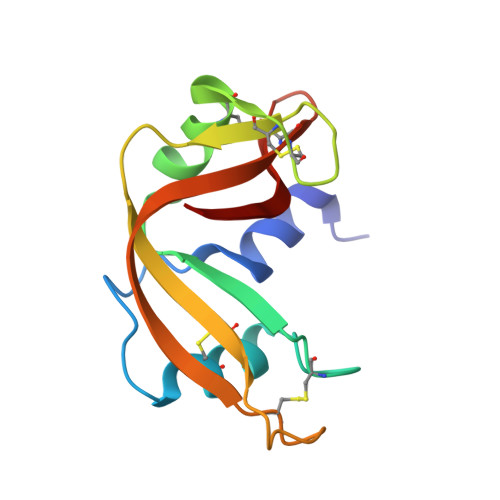
6PVU

RNase A in complex with hexametaphosphate


Experimental Data Snapshot

  • Method: X-RAY DIFFRACTION
  • Resolution: 1.49 Å
  • R-Value Free: 0.236 
  • R-Value Work: 0.202 
  • R-Value Observed: 0.203 

Starting Model: experimental
View more details

wwPDB Validation   3D Report Full Report

Currently 6PVU does not have a validation slider image.


This is version 1.4 of the entry. See complete history


Literature

Nucleoside Tetra- and Pentaphosphates Prepared Using a Tetraphosphorylation Reagent Are Potent Inhibitors of Ribonuclease A.

Shepard, S.M.Windsor, I.W.Raines, R.T.Cummins, C.C.

(2019) J Am Chem Soc 141: 18400-18404

  • DOI: https://doi.org/10.1021/jacs.9b09760
  • Primary Citation of Related Structures:  
    6PVU, 6PVV, 6PVW, 6PVX

  • PubMed Abstract: 

    Adenosine and uridine 5'-tetra- and 5'-pentaphosphates were synthesized from an activated tetrametaphosphate ([PPN] 2 [P 4 O 11 ], [PPN] 2 [ 1 ], PPN = bis(triphenylphosphine)iminium) and subsequently tested for inhibition of the enzymatic activity of ribonuclease A (RNase A). Reagent [PPN] 2 [ 1 ] reacts with unprotected uridine and adenosine in the presence of a base under anhydrous conditions to give nucleoside tetrametaphosphates. Ring opening of these intermediates with tetrabutylammonium hydroxide ([TBA][OH]) yields adenosine and uridine tetraphosphates ( p 4 A , p 4 U ) in 92% and 85% yields, respectively, from the starting nucleoside. Treatment of ([PPN] 2 [ 1 ]) with AMP or UMP yields nucleoside-monophosphate tetrametaphosphates ( cp 4 pA, cp 4 pU ) having limited aqueous stability. Ring opening of these ultraphosphates with [TBA][OH] yields p 5 A and p 5 U in 58% and 70% yield from AMP and UMP, respectively. We characterized inorganic and nucleoside-conjugated linear and cyclic oligophosphates as competitive inhibitors of RNase A. Increasing the chain length in both linear and cyclic inorganic oligophosphates resulted in improved binding affinity. Increasing the length of oligophosphates on the 5' position of adenosine beyond three had a deleterious effect on binding. Conversely, uridine nucleotides bearing 5' oligophosphates saw progressive increases in binding with chain length. We solved X-ray cocrystal structures of the highest affinity binders from several classes. The terminal phosphate of p 5 A binds in the P 1 enzymic subsite and forces the oligophosphate to adopt a convoluted conformation, while the oligophosphate of p 5 U binds in several extended conformations, targeting multiple cationic regions of the active-site cleft.


  • Organizational Affiliation

    Department of Chemistry , Massachusetts Institute of Technology , Cambridge Massachusetts 02139 , United States.


Macromolecules
Find similar proteins by:  (by identity cutoff)  |  3D Structure
Entity ID: 1
MoleculeChains Sequence LengthOrganismDetailsImage
Ribonuclease pancreatic
A, B
124Bos taurusMutation(s): 0 
EC: 4.6.1.18
UniProt
Find proteins for P61823 (Bos taurus)
Explore P61823 
Go to UniProtKB:  P61823
Entity Groups  
Sequence Clusters30% Identity50% Identity70% Identity90% Identity95% Identity100% Identity
UniProt GroupP61823
Sequence Annotations
Expand
  • Reference Sequence
Small Molecules
Ligands 1 Unique
IDChains Name / Formula / InChI Key2D Diagram3D Interactions
P0S (Subject of Investigation/LOI)
Query on P0S

Download Ideal Coordinates CCD File 
C [auth A],
D [auth B]
2,4,6,8,10,12-hexahydroxy-2lambda~5~,4lambda~5~,6lambda~5~,8lambda~5~,10lambda~5~,12lambda~5~-cyclohexaphosphoxane-2,4,6,8,10,12-hexone
H6 O18 P6
SZGVJLCXTSBVKL-UHFFFAOYSA-N
Experimental Data & Validation

Experimental Data

  • Method: X-RAY DIFFRACTION
  • Resolution: 1.49 Å
  • R-Value Free: 0.236 
  • R-Value Work: 0.202 
  • R-Value Observed: 0.203 
  • Space Group: C 1 2 1
Unit Cell:
Length ( Å )Angle ( ˚ )
a = 100.35α = 90
b = 32.64β = 90.65
c = 72.61γ = 90
Software Package:
Software NamePurpose
PHENIXrefinement
PDB_EXTRACTdata extraction
SAINTdata reduction
SADABSdata scaling
PHASERphasing

Structure Validation

View Full Validation Report

Currently 6PVU does not have a validation slider image.


Entry History & Funding Information

Deposition Data


Funding OrganizationLocationGrant Number
National Institutes of Health/National Cancer Institute (NIH/NCI)United StatesR01 CA073808

Revision History  (Full details and data files)

  • Version 1.0: 2019-11-06
    Type: Initial release
  • Version 1.1: 2019-11-13
    Changes: Data collection, Database references
  • Version 1.2: 2019-12-04
    Changes: Author supporting evidence, Database references
  • Version 1.3: 2023-10-11
    Changes: Data collection, Database references, Refinement description
  • Version 1.4: 2024-10-30
    Changes: Structure summary