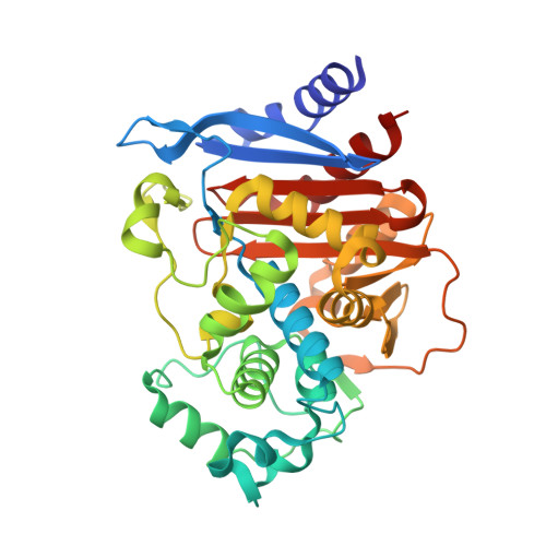
Structural Insights into Inhibition of the Acinetobacter-Derived Cephalosporinase ADC-7 by Ceftazidime and Its Boronic Acid Transition State Analog.
Curtis, B.N., Smolen, K.A., Barlow, S.J., Caselli, E., Prati, F., Taracila, M.A., Bonomo, R.A., Wallar, B.J., Powers, R.A.(2020) Antimicrob Agents Chemother 64
- PubMed: 32988830
- DOI: https://doi.org/10.1128/AAC.01183-20
- Primary Citation of Related Structures:
6PWL, 6PWM - PubMed Abstract:
Extended-spectrum class C β-lactamases have evolved to rapidly inactivate expanded-spectrum cephalosporins, a class of antibiotics designed to be resistant to hydrolysis by β-lactamase enzymes. To better understand the mechanism by which A cinetobacter- d erived c ephalosporinase-7 (ADC-7), a chromosomal AmpC enzyme, hydrolyzes these molecules, we determined the X-ray crystal structure of ADC-7 in an acyl-enzyme complex with the cephalosporin ceftazidime (2.40 Å) as well as in complex with a boronic acid transition state analog inhibitor that contains the R1 side chain of ceftazidime (1.67 Å). In the acyl-enzyme complex, the carbonyl oxygen is situated in the oxyanion hole where it makes key stabilizing interactions with the main chain nitrogens of Ser64 and Ser315. The boronic acid O1 hydroxyl group is similarly positioned in this area. Conserved residues Gln120 and Asn152 form hydrogen bonds with the amide group of the R1 side chain in both complexes. These complexes represent two steps in the hydrolysis of expanded-spectrum cephalosporins by ADC-7 and offer insight into the inhibition of ADC-7 by ceftazidime through displacement of the deacylating water molecule as well as blocking its trajectory to the acyl carbonyl carbon. In addition, the transition state analog inhibitor, LP06, was shown to bind with high affinity to ADC-7 ( K i , 50 nM) and was able to restore ceftazidime susceptibility, offering the potential for optimization efforts of this type of inhibitor.
Organizational Affiliation:
Department of Chemistry, Grand Valley State University, Allendale, Michigan, USA.


