6R4T

Crystal structure of the Pri1 subunit of human primase bound to vidarabine triphosphate


Experimental Data Snapshot

  • Method: X-RAY DIFFRACTION
  • Resolution: 2.35 Å
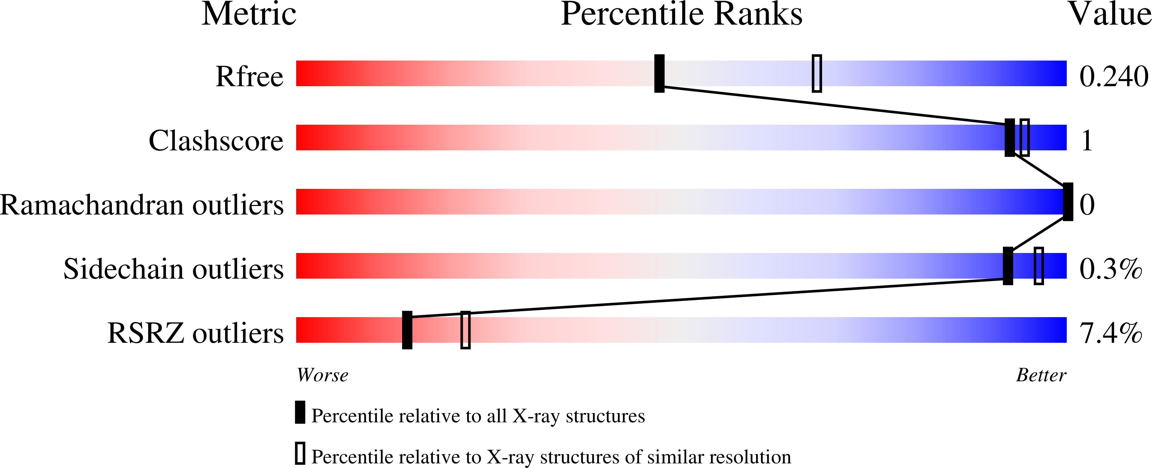
  • R-Value Free: 0.239 
  • R-Value Work: 0.209 
  • R-Value Observed: 0.211 

Starting Model: experimental
View more details

wwPDB Validation   3D Report Full Report


Ligand Structure Quality Assessment 


This is version 1.2 of the entry. See complete history


Literature

Structural Basis for Inhibition of Human Primase by Arabinofuranosyl Nucleoside Analogues Fludarabine and Vidarabine.

Holzer, S.Rzechorzek, N.J.Short, I.R.Jenkyn-Bedford, M.Pellegrini, L.Kilkenny, M.L.

(2019) ACS Chem Biol 14: 1904-1912

  • DOI: https://doi.org/10.1021/acschembio.9b00367
  • Primary Citation of Related Structures:  
    6R4S, 6R4T, 6R4U, 6R5D, 6R5E, 6RB4

  • PubMed Abstract: 

    Nucleoside analogues are widely used in clinical practice as chemotherapy drugs. Arabinose nucleoside derivatives such as fludarabine are effective in the treatment of patients with acute and chronic leukemias and non-Hodgkin's lymphomas. Although nucleoside analogues are generally known to function by inhibiting DNA synthesis in rapidly proliferating cells, the identity of their in vivo targets and mechanism of action are often not known in molecular detail. Here we provide a structural basis for arabinose nucleotide-mediated inhibition of human primase, the DNA-dependent RNA polymerase responsible for initiation of DNA synthesis in DNA replication. Our data suggest ways in which the chemical structure of fludarabine could be modified to improve its specificity and affinity toward primase, possibly leading to less toxic and more effective therapeutic agents.


  • Organizational Affiliation

    Department of Biochemistry , University of Cambridge , 80 Tennis Court Road , Cambridge CB2 1GA , U.K.


Macromolecules
Find similar proteins by:  (by identity cutoff)  |  3D Structure
Entity ID: 1
MoleculeChains Sequence LengthOrganismDetailsImage
DNA primase small subunitA,
B [auth D]
410Homo sapiensMutation(s): 0 
Gene Names: PRIM1
EC: 2.7.7 (PDB Primary Data), 2.7.7.102 (UniProt)
UniProt & NIH Common Fund Data Resources
Find proteins for P49642 (Homo sapiens)
Explore P49642 
Go to UniProtKB:  P49642
PHAROS:  P49642
GTEx:  ENSG00000198056 
Entity Groups  
Sequence Clusters30% Identity50% Identity70% Identity90% Identity95% Identity100% Identity
UniProt GroupP49642
Sequence Annotations
Expand
  • Reference Sequence
Small Molecules
Ligands 4 Unique
IDChains Name / Formula / InChI Key2D Diagram3D Interactions
HEJ (Subject of Investigation/LOI)
Query on HEJ

Download Ideal Coordinates CCD File 
E [auth A],
J [auth D]
9-{5-O-[(S)-hydroxy{[(R)-hydroxy(phosphonooxy)phosphoryl]oxy}phosphoryl]-beta-D-arabinofuranosyl}-9H-purin-6-amine
C10 H16 N5 O13 P3
ZKHQWZAMYRWXGA-UHTZMRCNSA-N
ZN
Query on ZN

Download Ideal Coordinates CCD File 
C [auth A],
H [auth D]
ZINC ION
Zn
PTFCDOFLOPIGGS-UHFFFAOYSA-N
EDO
Query on EDO

Download Ideal Coordinates CCD File 
F [auth A],
G [auth A]
1,2-ETHANEDIOL
C2 H6 O2
LYCAIKOWRPUZTN-UHFFFAOYSA-N
MN
Query on MN

Download Ideal Coordinates CCD File 
D [auth A],
I [auth D]
MANGANESE (II) ION
Mn
WAEMQWOKJMHJLA-UHFFFAOYSA-N
Experimental Data & Validation

Experimental Data

  • Method: X-RAY DIFFRACTION
  • Resolution: 2.35 Å
  • R-Value Free: 0.239 
  • R-Value Work: 0.209 
  • R-Value Observed: 0.211 
  • Space Group: C 2 2 21
Unit Cell:
Length ( Å )Angle ( ˚ )
a = 111.09α = 90
b = 118.97β = 90
c = 150.72γ = 90
Software Package:
Software NamePurpose
PHENIXrefinement
XDSdata reduction
XDSdata scaling
Cootmodel building
PHENIXphasing

Structure Validation

View Full Validation Report


Ligand Structure Quality Assessment 


Entry History & Funding Information

Deposition Data


Funding OrganizationLocationGrant Number
Wellcome TrustUnited Kingdom104641/Z/14/Z

Revision History  (Full details and data files)

  • Version 1.0: 2019-09-11
    Type: Initial release
  • Version 1.1: 2019-10-02
    Changes: Data collection, Database references
  • Version 1.2: 2024-01-24
    Changes: Data collection, Database references, Derived calculations, Refinement description