6R7R

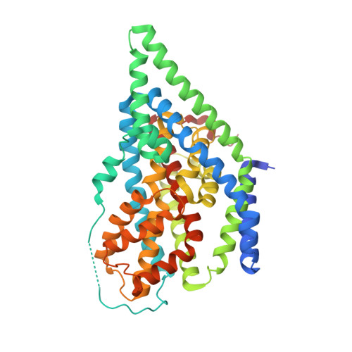
Crystal structure of the glutamate transporter homologue GltTk in complex with D-aspartate


Experimental Data Snapshot

  • Method: X-RAY DIFFRACTION
  • Resolution: 2.80 Å
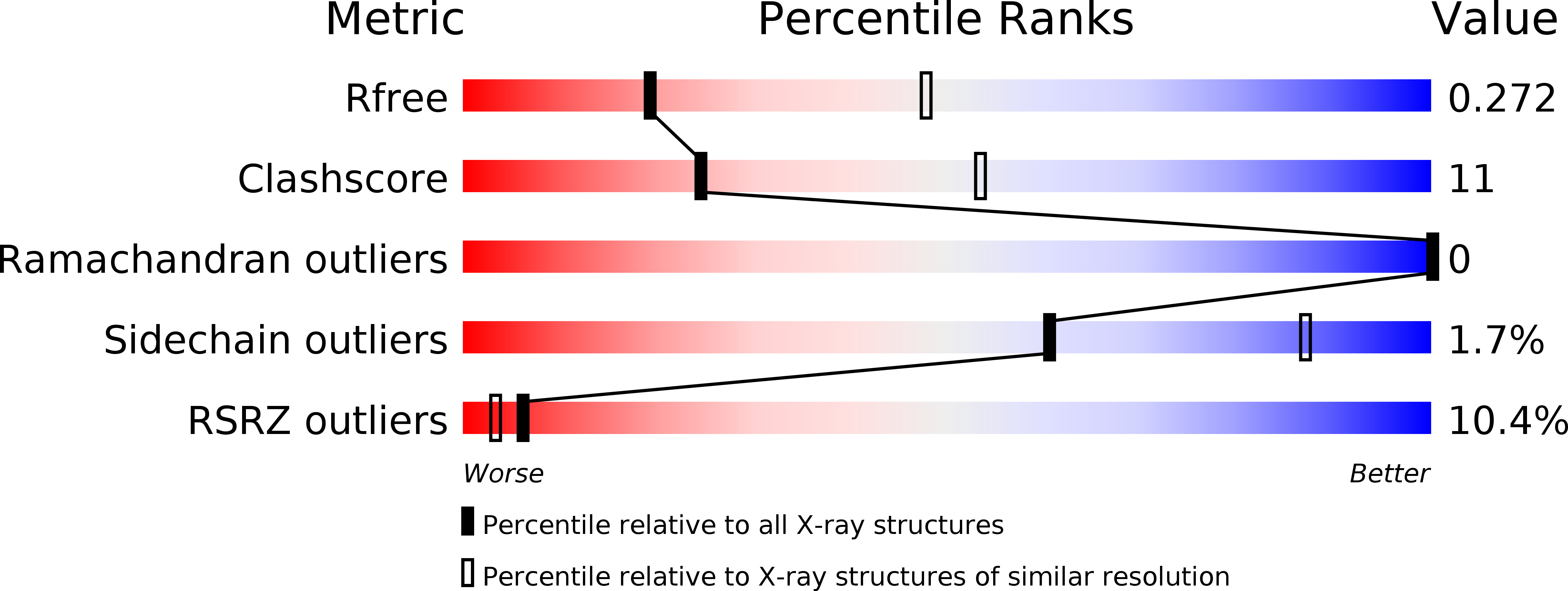
  • R-Value Free: 0.269 
  • R-Value Work: 0.232 
  • R-Value Observed: 0.234 

Starting Model: experimental
View more details

wwPDB Validation   3D Report Full Report


Ligand Structure Quality Assessment 


This is version 1.3 of the entry. See complete history


Literature

Binding and transport of D-aspartate by the glutamate transporter homolog Glt Tk .

Arkhipova, V.Trinco, G.Ettema, T.W.Jensen, S.Slotboom, D.J.Guskov, A.

(2019) Elife 8

  • DOI: https://doi.org/10.7554/eLife.45286
  • Primary Citation of Related Structures:  
    6R7R

  • PubMed Abstract: 

    Mammalian glutamate transporters are crucial players in neuronal communication as they perform neurotransmitter reuptake from the synaptic cleft. Besides L-glutamate and L-aspartate, they also recognize D-aspartate, which might participate in mammalian neurotransmission and/or neuromodulation. Much of the mechanistic insight in glutamate transport comes from studies of the archeal homologs Glt Ph from Pyrococcus horikoshii and Glt Tk from Thermococcus kodakarensis . Here, we show that Glt Tk transports D-aspartate with identical Na + : substrate coupling stoichiometry as L-aspartate, and that the affinities ( K d and K m ) for the two substrates are similar. We determined a crystal structure of Glt Tk with bound D-aspartate at 2.8 Å resolution. Comparison of the L- and D-aspartate bound Glt Tk structures revealed that D-aspartate is accommodated with only minor rearrangements in the structure of the binding site. The structure explains how the geometrically different molecules L- and D-aspartate are recognized and transported by the protein in the same way.


  • Organizational Affiliation

    Groningen Biomolecular Sciences and Biotechnology Institute, Zernike Institute for Advanced Materials, University of Groningen, Groningen, The Netherlands.


Macromolecules
Find similar proteins by:  (by identity cutoff)  |  3D Structure
Entity ID: 1
MoleculeChains Sequence LengthOrganismDetailsImage
Proton/glutamate symporter, SDF family
A, B, C
438Thermococcus kodakarensis KOD1Mutation(s): 0 
Gene Names: TK0986
Membrane Entity: Yes 
UniProt
Find proteins for Q5JID0 (Thermococcus kodakarensis (strain ATCC BAA-918 / JCM 12380 / KOD1))
Explore Q5JID0 
Go to UniProtKB:  Q5JID0
Entity Groups  
Sequence Clusters30% Identity50% Identity70% Identity90% Identity95% Identity100% Identity
UniProt GroupQ5JID0
Sequence Annotations
Expand
  • Reference Sequence
Small Molecules
Ligands 8 Unique
IDChains Name / Formula / InChI Key2D Diagram3D Interactions
DMU
Query on DMU

Download Ideal Coordinates CCD File 
L [auth A]DECYL-BETA-D-MALTOPYRANOSIDE
C22 H42 O11
WOQQAWHSKSSAGF-WXFJLFHKSA-N
P6G
Query on P6G

Download Ideal Coordinates CCD File 
V [auth B],
W [auth B]
HEXAETHYLENE GLYCOL
C12 H26 O7
IIRDTKBZINWQAW-UHFFFAOYSA-N
1PE
Query on 1PE

Download Ideal Coordinates CCD File 
Z [auth B]PENTAETHYLENE GLYCOL
C10 H22 O6
JLFNLZLINWHATN-UHFFFAOYSA-N
PG4
Query on PG4

Download Ideal Coordinates CCD File 
K [auth A]TETRAETHYLENE GLYCOL
C8 H18 O5
UWHCKJMYHZGTIT-UHFFFAOYSA-N
PGE
Query on PGE

Download Ideal Coordinates CCD File 
M [auth A],
X [auth B],
Y [auth B]
TRIETHYLENE GLYCOL
C6 H14 O4
ZIBGPFATKBEMQZ-UHFFFAOYSA-N
DAS (Subject of Investigation/LOI)
Query on DAS

Download Ideal Coordinates CCD File 
DA [auth C],
G [auth A],
Q [auth B]
D-ASPARTIC ACID
C4 H7 N O4
CKLJMWTZIZZHCS-UWTATZPHSA-N
PEG
Query on PEG

Download Ideal Coordinates CCD File 
EA [auth C]
FA [auth C]
GA [auth C]
H [auth A]
HA [auth C]
EA [auth C],
FA [auth C],
GA [auth C],
H [auth A],
HA [auth C],
I [auth A],
IA [auth C],
J [auth A],
R [auth B],
S [auth B],
T [auth B],
U [auth B]
DI(HYDROXYETHYL)ETHER
C4 H10 O3
MTHSVFCYNBDYFN-UHFFFAOYSA-N
NA
Query on NA

Download Ideal Coordinates CCD File 
AA [auth C]
BA [auth C]
CA [auth C]
D [auth A]
E [auth A]
AA [auth C],
BA [auth C],
CA [auth C],
D [auth A],
E [auth A],
F [auth A],
N [auth B],
O [auth B],
P [auth B]
SODIUM ION
Na
FKNQFGJONOIPTF-UHFFFAOYSA-N
Experimental Data & Validation

Experimental Data

  • Method: X-RAY DIFFRACTION
  • Resolution: 2.80 Å
  • R-Value Free: 0.269 
  • R-Value Work: 0.232 
  • R-Value Observed: 0.234 
  • Space Group: P 32 2 1
Unit Cell:
Length ( Å )Angle ( ˚ )
a = 116.55α = 90
b = 116.55β = 90
c = 314.77γ = 120
Software Package:
Software NamePurpose
PHENIXrefinement
XDSdata reduction
XSCALEdata scaling
PHASERphasing

Structure Validation

View Full Validation Report


Ligand Structure Quality Assessment 


Entry History & Funding Information

Deposition Data


Funding OrganizationLocationGrant Number
Netherlands Organisation for Scientific ResearchNetherlands865.11.001

Revision History  (Full details and data files)

  • Version 1.0: 2019-04-17
    Type: Initial release
  • Version 1.1: 2019-05-01
    Changes: Data collection, Database references
  • Version 1.2: 2020-09-30
    Changes: Database references
  • Version 1.3: 2024-01-24
    Changes: Data collection, Database references, Refinement description