6T0Q

Pleurotus Ostreatus Lectin (POL), apo form


Experimental Data Snapshot

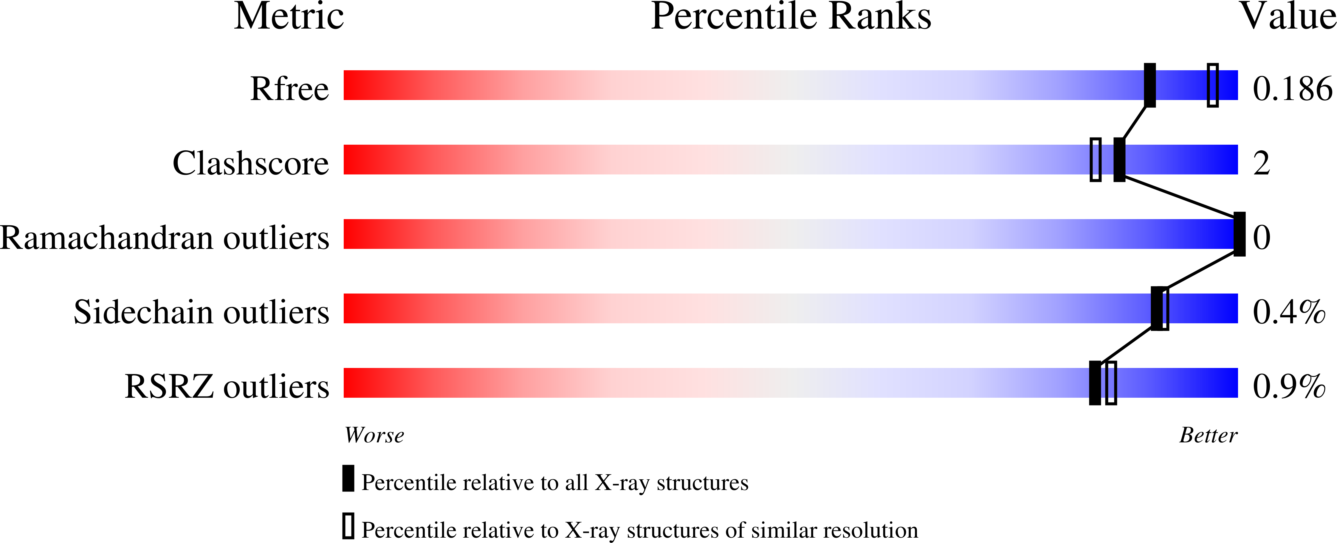
  • Method: X-RAY DIFFRACTION
  • Resolution: 2.05 Å
  • R-Value Free: 0.187 
  • R-Value Work: 0.172 
  • R-Value Observed: 0.173 

wwPDB Validation   3D Report Full Report


This is version 3.0 of the entry. See complete history


Literature

Structure and properties of the oyster mushroom (Pleurotus ostreatus) lectin.

Perduca, M.Destefanis, L.Bovi, M.Galliano, M.Munari, F.Assfalg, M.Ferrari, F.Monaco, H.L.Capaldi, S.

(2020) Glycobiology 30: 550-562

  • DOI: https://doi.org/10.1093/glycob/cwaa006
  • Primary Citation of Related Structures:  
    6T0Q, 6T1D

  • PubMed Abstract: 

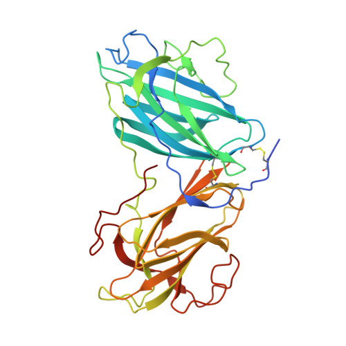
    Pleurotus ostreatus Lectin (POL) is a 353 amino acid chain lectin that can be purified from the fruiting bodies of the very well-known and widely diffused edible oyster mushrooms (P. ostreatus). The lectin has been partially characterized by different groups and, although it was crystallized about 20 years ago, its 3D structure and the details of its interactions with carbohydrates are still unknown. This paper reports the 3D structure and ligand-binding properties of POL. We have determined the X-ray structure of the apo-protein purified from the fruiting bodies of the mushroom and that of the recombinant protein in complex with melibiose to a resolution of about 2 Å. The lectin is a homodimer in which the two polypeptide chains are linked by a disulfide bridge. A POL monomer is composed of two highly homologous β-jellyroll domains each of which containing a calcium-dependent carbohydrate-binding site. A high degree of sequence similarity is observed between the two carbohydrate-binding modules present in each monomer. The structure of the lectin in complex with melibiose reveals that a POL dimer has four calcium-dependent carbohydrate-binding sites. The interaction with sugars in solution has been characterized by isothermal titration calorimetry and saturation transfer difference NMR and it sheds new light on the molecular determinants of POL specificity. The lectin exhibits in vitro antiproliferative effects against human cancer cell lines and presents structural similarity with the prototype member of the CBM67 family, the noncatalytic domain of Streptomyces avermitilis α-rhamnosidase.


  • Organizational Affiliation

    Biocrystallography and Nanostructure Laboratory, Department of Biotechnology, University of Verona, Strada Le Grazie 15, 37134 Verona, Italy.


Macromolecules
Find similar proteins by:  (by identity cutoff)  |  3D Structure
Entity ID: 1
MoleculeChains Sequence LengthOrganismDetailsImage
Lectin353Pleurotus ostreatusMutation(s): 0 
UniProt
Find proteins for E7E2M2 (Pleurotus ostreatus)
Explore E7E2M2 
Go to UniProtKB:  E7E2M2
Entity Groups  
Sequence Clusters30% Identity50% Identity70% Identity90% Identity95% Identity100% Identity
UniProt GroupE7E2M2
Sequence Annotations
Expand
  • Reference Sequence
Oligosaccharides

Help

Entity ID: 2
MoleculeChains Length2D Diagram Glycosylation3D Interactions
2-acetamido-2-deoxy-beta-D-glucopyranose-(1-4)-2-acetamido-2-deoxy-beta-D-glucopyranoseB [auth C],
C [auth D]
2N/A
Glycosylation Resources
GlyTouCan:  G42666HT
GlyCosmos:  G42666HT
GlyGen:  G42666HT
Small Molecules
Ligands 3 Unique
IDChains Name / Formula / InChI Key2D Diagram3D Interactions
NAG
Query on NAG

Download Ideal Coordinates CCD File 
D [auth A],
E [auth A]
2-acetamido-2-deoxy-beta-D-glucopyranose
C8 H15 N O6
OVRNDRQMDRJTHS-FMDGEEDCSA-N
GOL
Query on GOL

Download Ideal Coordinates CCD File 
H [auth A],
I [auth A],
J [auth A],
K [auth A]
GLYCEROL
C3 H8 O3
PEDCQBHIVMGVHV-UHFFFAOYSA-N
CA (Subject of Investigation/LOI)
Query on CA

Download Ideal Coordinates CCD File 
F [auth A],
G [auth A]
CALCIUM ION
Ca
BHPQYMZQTOCNFJ-UHFFFAOYSA-N
Experimental Data & Validation

Experimental Data

  • Method: X-RAY DIFFRACTION
  • Resolution: 2.05 Å
  • R-Value Free: 0.187 
  • R-Value Work: 0.172 
  • R-Value Observed: 0.173 
  • Space Group: P 61 2 2
Unit Cell:
Length ( Å )Angle ( ˚ )
a = 153.07α = 90
b = 153.07β = 90
c = 145.811γ = 120
Software Package:
Software NamePurpose
PHENIXrefinement
MOSFLMdata reduction
SCALAdata scaling
PDB_EXTRACTdata extraction
SHARPphasing

Structure Validation

View Full Validation Report


Entry History 

Deposition Data

Revision History  (Full details and data files)

  • Version 1.0: 2020-02-19
    Type: Initial release
  • Version 2.0: 2020-07-29
    Type: Remediation
    Reason: Carbohydrate remediation
    Changes: Advisory, Atomic model, Data collection, Database references, Derived calculations, Structure summary
  • Version 3.0: 2022-12-07
    Changes: Advisory, Atomic model, Data collection, Database references, Derived calculations, Structure summary