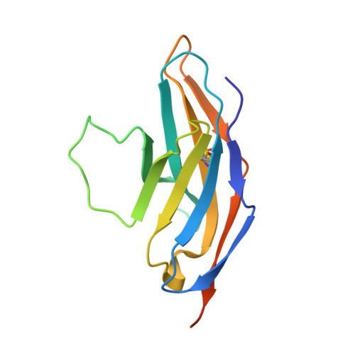
A high-affinity human PD-1/PD-L2 complex informs avenues for small-molecule immune checkpoint drug discovery.
Tang, S., Kim, P.S.(2019) Proc Natl Acad Sci U S A 116: 24500-24506
- PubMed: 31727844
- DOI: https://doi.org/10.1073/pnas.1916916116
- Primary Citation of Related Structures:
6UMT, 6UMU, 6UMV - PubMed Abstract:
Immune checkpoint blockade of programmed death-1 (PD-1) by monoclonal antibody drugs has delivered breakthroughs in the treatment of cancer. Nonetheless, small-molecule PD-1 inhibitors could lead to increases in treatment efficacy, safety, and global access. While the ligand-binding surface of apo-PD-1 is relatively flat, it harbors a striking pocket in the murine PD-1/PD-L2 structure. An analogous pocket in human PD-1 may serve as a small-molecule drug target, but the structure of the human complex is unknown. Because the CC' and FG loops in murine PD-1 adopt new conformations upon binding PD-L2, we hypothesized that mutations in these two loops could be coupled to pocket formation and alter PD-1's affinity for PD-L2. Here, we conducted deep mutational scanning in these loops and used yeast surface display to select for enhanced PD-L2 binding. A PD-1 variant with three substitutions binds PD-L2 with an affinity two orders of magnitude higher than that of the wild-type protein, permitting crystallization of the complex. We determined the X-ray crystal structures of the human triple-mutant PD-1/PD-L2 complex and the apo triple-mutant PD-1 variant at 2.0 Å and 1.2 Å resolution, respectively. Binding of PD-L2 is accompanied by formation of a prominent pocket in human PD-1, as well as substantial conformational changes in the CC' and FG loops. The structure of the apo triple-mutant PD-1 shows that the CC' loop adopts the ligand-bound conformation, providing support for allostery between the loop and pocket. This human PD-1/PD-L2 structure provide critical insights for the design and discovery of small-molecule PD-1 inhibitors.
Organizational Affiliation:
Stanford ChEM-H, Stanford University, Stanford, CA 94305.


