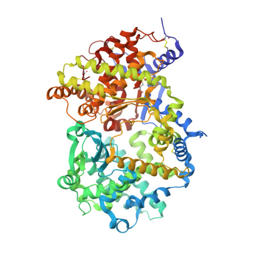
6XVP

Crystal structure of Neprilysin in complex with Sampatrilat.


Experimental Data Snapshot

  • Method: X-RAY DIFFRACTION
  • Resolution: 2.65 Å
  • R-Value Free: 0.237 
  • R-Value Work: 0.191 
  • R-Value Observed: 0.194 

Starting Model: experimental
View more details

wwPDB Validation   3D Report Full Report


Ligand Structure Quality Assessment 


This is version 1.5 of the entry. See complete history


Literature

Molecular Basis for Omapatrilat and Sampatrilat Binding to Neprilysin-Implications for Dual Inhibitor Design with Angiotensin-Converting Enzyme.

Sharma, U.Cozier, G.E.Sturrock, E.D.Acharya, K.R.

(2020) J Med Chem 63: 5488-5500

  • DOI: https://doi.org/10.1021/acs.jmedchem.0c00441
  • Primary Citation of Related Structures:  
    6SUK, 6SVY, 6XVP

  • PubMed Abstract: 

    Neprilysin (NEP) and angiotensin-converting enzyme (ACE) are two key zinc-dependent metallopeptidases in the natriuretic peptide and kinin systems and renin-angiotensin-aldosterone system, respectively. They play an important role in blood pressure regulation and reducing the risk of heart failure. Vasopeptidase inhibitors omapatrilat and sampatrilat possess dual activity against these enzymes by blocking the ACE-dependent conversion of angiotensin I to the potent vasoconstrictor angiotensin II while simultaneously halting the NEP-dependent degradation of vasodilator atrial natriuretic peptide. Here, we report crystal structures of omapatrilat, sampatrilat, and sampatrilat-ASP (a sampatrilat analogue) in complex with NEP at 1.75, 2.65, and 2.6 Å, respectively. A detailed analysis of these structures and the corresponding structures of ACE with these inhibitors has provided the molecular basis of dual inhibitor recognition involving the catalytic site in both enzymes. This new information will be very useful in the design of safer and more selective vasopeptidase inhibitors of NEP and ACE for effective treatment in hypertension and heart failure.


  • Organizational Affiliation

    Department of Biology and Biochemistry, University of Bath, Claverton Down, Bath BA2 7AY, United Kingdom.


Macromolecules
Find similar proteins by:  (by identity cutoff)  |  3D Structure
Entity ID: 1
MoleculeChains Sequence LengthOrganismDetailsImage
Neprilysin699Homo sapiensMutation(s): 0 
Gene Names: MMEEPN
EC: 3.4.24.11
UniProt & NIH Common Fund Data Resources
Find proteins for P08473 (Homo sapiens)
Explore P08473 
Go to UniProtKB:  P08473
PHAROS:  P08473
GTEx:  ENSG00000196549 
Entity Groups  
Sequence Clusters30% Identity50% Identity70% Identity90% Identity95% Identity100% Identity
UniProt GroupP08473
Glycosylation
Glycosylation Sites: 4Go to GlyGen: P08473-1
Sequence Annotations
Expand
  • Reference Sequence
Small Molecules
Ligands 4 Unique
IDChains Name / Formula / InChI Key2D Diagram3D Interactions
D0Z (Subject of Investigation/LOI)
Query on D0Z

Download Ideal Coordinates CCD File 
C [auth A]Sampatrilat
C26 H40 N4 O9 S
LPUDGHQMOAHMMF-JBACZVJFSA-N
NAG
Query on NAG

Download Ideal Coordinates CCD File 
D [auth A],
E [auth A],
F [auth A],
G [auth A]
2-acetamido-2-deoxy-beta-D-glucopyranose
C8 H15 N O6
OVRNDRQMDRJTHS-FMDGEEDCSA-N
ZN
Query on ZN

Download Ideal Coordinates CCD File 
B [auth A]ZINC ION
Zn
PTFCDOFLOPIGGS-UHFFFAOYSA-N
EDO
Query on EDO

Download Ideal Coordinates CCD File 
H [auth A],
I [auth A],
J [auth A]
1,2-ETHANEDIOL
C2 H6 O2
LYCAIKOWRPUZTN-UHFFFAOYSA-N
Experimental Data & Validation

Experimental Data

  • Method: X-RAY DIFFRACTION
  • Resolution: 2.65 Å
  • R-Value Free: 0.237 
  • R-Value Work: 0.191 
  • R-Value Observed: 0.194 
  • Space Group: P 32 2 1
Unit Cell:
Length ( Å )Angle ( ˚ )
a = 108.089α = 90
b = 108.089β = 90
c = 112.826γ = 120
Software Package:
Software NamePurpose
PHENIXrefinement
PHENIXrefinement
DIALSdata reduction
Aimlessdata scaling
PHASERphasing

Structure Validation

View Full Validation Report


Ligand Structure Quality Assessment 


Entry History & Funding Information

Deposition Data


Funding OrganizationLocationGrant Number
Medical Research Council (MRC, United Kingdom)United KingdomMR/M026647/1

Revision History  (Full details and data files)

  • Version 1.0: 2020-05-13
    Type: Initial release
  • Version 1.1: 2020-05-20
    Changes: Database references
  • Version 1.2: 2020-06-10
    Changes: Database references
  • Version 1.3: 2020-07-29
    Type: Remediation
    Reason: Carbohydrate remediation
    Changes: Data collection, Derived calculations, Structure summary
  • Version 1.4: 2024-01-24
    Changes: Data collection, Database references, Refinement description, Structure summary
  • Version 1.5: 2024-10-23
    Changes: Structure summary