6YNF

GAPDH purified from the supernatant of HEK293F cells: crystal form 3 of 4.


Experimental Data Snapshot

  • Method: X-RAY DIFFRACTION
  • Resolution: 2.39 Å
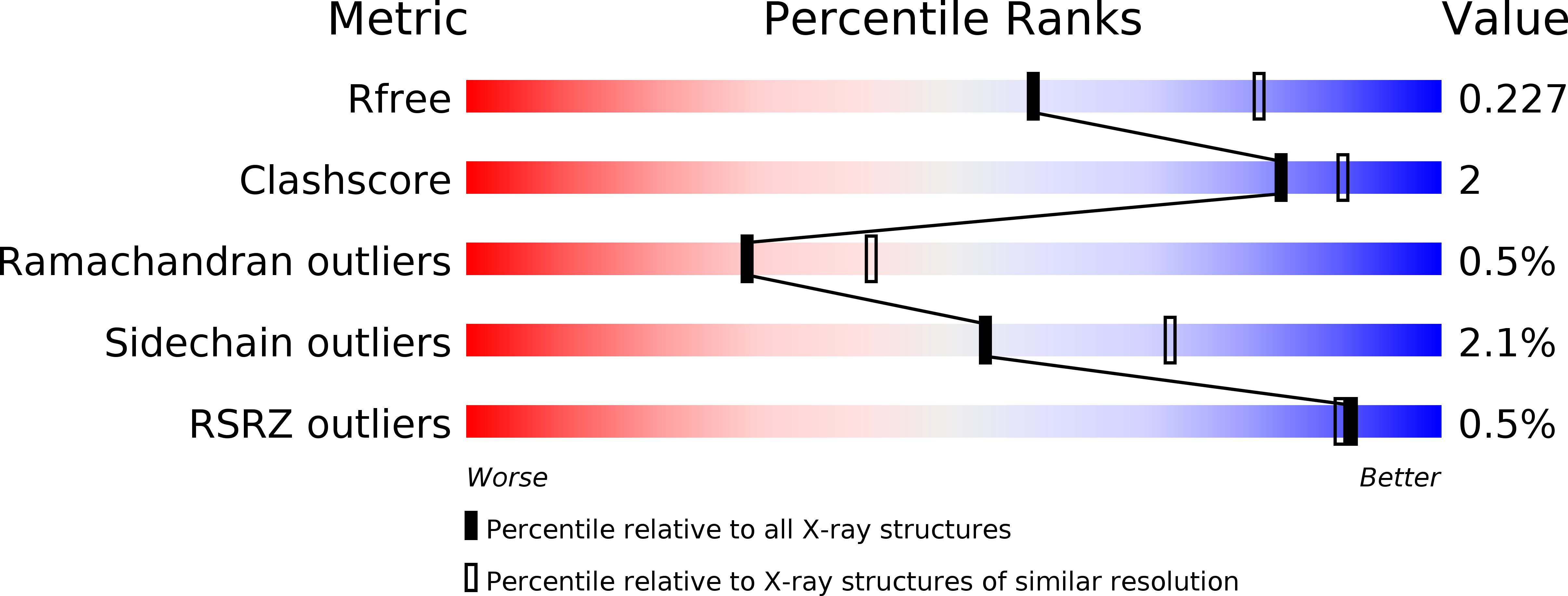
  • R-Value Free: 0.219 
  • R-Value Work: 0.179 
  • R-Value Observed: 0.181 

Starting Model: experimental
View more details

wwPDB Validation   3D Report Full Report


This is version 1.2 of the entry. See complete history


Literature

Partial catalytic Cys oxidation of human GAPDH to Cys-sulfonic acid.

Lia, A.Dowle, A.Taylor, C.Santino, A.Roversi, P.

(2020) Wellcome Open Res 5: 114-114

  • DOI: https://doi.org/10.12688/wellcomeopenres.15893.2
  • Primary Citation of Related Structures:  
    6YND, 6YNE, 6YNF, 6YNH

  • PubMed Abstract: 

    Background : n-Glyceraldehyde-3-phosphate dehydrogenase (GAPDH) catalyses the NAD + -dependent oxidative phosphorylation of n-glyceraldehyde-3-phosphate to 1,3-diphospho-n-glycerate and its reverse reaction in glycolysis and gluconeogenesis. Methods : Four distinct crystal structures of human n-Glyceraldehyde-3-phosphate dehydrogenase ( Hs GAPDH) have been determined from protein purified from the supernatant of HEK293F human epithelial kidney cells. Results : X-ray crystallography and mass-spectrometry indicate that the catalytic cysteine of the protein ( Hs GAPDH Cys152) is partially oxidised to cysteine S-sulfonic acid. The average occupancy for the Cys152-S-sulfonic acid modification over the 20 crystallographically independent copies of Hs GAPDH across three of the crystal forms obtained is 0.31±0.17. Conclusions : The modification induces no significant structural changes on the tetrameric enzyme, and only makes aspecific contacts to surface residues in the active site, in keeping with the hypothesis that the oxidising conditions of the secreted mammalian cell expression system result in Hs GAPDH catalytic cysteine S-sulfonic acid modification and irreversible inactivation of the enzyme.


  • Organizational Affiliation

    Leicester Institute of Chemical and Structural Biology and Department of Molecular and Cell Biology, University of Leicester, Henry Wellcome Building, Lancaster Road, LE1 7HB, UK.


Macromolecules
Find similar proteins by:  (by identity cutoff)  |  3D Structure
Entity ID: 1
MoleculeChains Sequence LengthOrganismDetailsImage
Glyceraldehyde-3-phosphate dehydrogenase
A, B, C, D, E
A, B, C, D, E, F, G, H
334Homo sapiensMutation(s): 0 
EC: 1.2.1.12 (PDB Primary Data), 2.6.99 (PDB Primary Data)
UniProt & NIH Common Fund Data Resources
Find proteins for P04406 (Homo sapiens)
Explore P04406 
Go to UniProtKB:  P04406
PHAROS:  P04406
GTEx:  ENSG00000111640 
Entity Groups  
Sequence Clusters30% Identity50% Identity70% Identity90% Identity95% Identity100% Identity
UniProt GroupP04406
Sequence Annotations
Expand
  • Reference Sequence
Small Molecules
Experimental Data & Validation

Experimental Data

  • Method: X-RAY DIFFRACTION
  • Resolution: 2.39 Å
  • R-Value Free: 0.219 
  • R-Value Work: 0.179 
  • R-Value Observed: 0.181 
  • Space Group: P 1 21 1
Unit Cell:
Length ( Å )Angle ( ˚ )
a = 87.136α = 90
b = 111.429β = 96.02
c = 135.943γ = 90
Software Package:
Software NamePurpose
BUSTERrefinement
XDSdata reduction
Aimlessdata scaling
MOLREPphasing

Structure Validation

View Full Validation Report


Entry History & Funding Information

Deposition Data


Funding OrganizationLocationGrant Number
Wellcome TrustUnited Kingdom204801/Z/16/Z
Wellcome TrustUnited Kingdom214090/Z/18/Z

Revision History  (Full details and data files)

  • Version 1.0: 2020-05-06
    Type: Initial release
  • Version 1.1: 2020-10-14
    Changes: Database references
  • Version 1.2: 2024-01-24
    Changes: Data collection, Database references, Refinement description