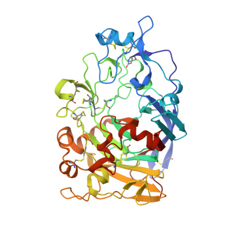
6YOZ

HiCel7B labelled with b-1,4-glucosyl cyclophellitol


Experimental Data Snapshot

  • Method: X-RAY DIFFRACTION
  • Resolution: 1.88 Å
  • R-Value Free: 0.236 
  • R-Value Work: 0.202 

Starting Model: experimental
View more details

wwPDB Validation   3D Report Full Report


Ligand Structure Quality Assessment 


This is version 1.4 of the entry. See complete history


Literature

Glycosylated cyclophellitol-derived activity-based probes and inhibitors for cellulases.

de Boer, C.McGregor, N.G.S.Peterse, E.Schroder, S.P.Florea, B.I.Jiang, J.Reijngoud, J.Ram, A.F.J.van Wezel, G.P.van der Marel, G.A.Codee, J.D.C.Overkleeft, H.S.Davies, G.J.

(2020) RSC Chem Biol 1: 148-155

  • DOI: https://doi.org/10.1039/d0cb00045k
  • Primary Citation of Related Structures:  
    6YOZ, 6YP1

  • PubMed Abstract: 

    Cellulases and related β-1,4-glucanases are essential components of lignocellulose-degrading enzyme mixtures. The detection of β-1,4-glucanase activity typically relies on monitoring the breakdown of purified lignocellulose-derived substrates or synthetic chromogenic substrates, limiting the activities which can be detected and complicating the tracing of activity back to specific components within complex enzyme mixtures. As a tool for the rapid detection and identification of β-1,4-glucanases, a series of glycosylated cyclophellitol inhibitors mimicking β-1,4-glucan oligosaccharides have been synthesised. These compounds are highly efficient inhibitors of HiCel7B, a well-known GH7 endo -β-1,4-glucanase. An elaborated activity-based probe facilitated the direct detection and identification of β-1,4-glucanases within a complex fungal secretome without any detectable cross-reactivity with β-d-glucosidases. These probes and inhibitors add valuable new capacity to the growing toolbox of cyclophellitol-derived probes for the activity-based profiling of biomass-degrading enzymes.


  • Organizational Affiliation

    Leiden Institute of Chemistry, Leiden University Einsteinweg 55 2300 RA Leiden The Netherlands [email protected].


Macromolecules
Find similar proteins by:  (by identity cutoff)  |  3D Structure
Entity ID: 1
MoleculeChains Sequence LengthOrganismDetailsImage
Endoglucanase 1A [auth AAA],
B [auth BBB]
400Mycothermus thermophilusMutation(s): 0 
Gene Names: CEL7B
EC: 3.2.1.4
UniProt
Find proteins for P56680 (Humicola insolens)
Explore P56680 
Go to UniProtKB:  P56680
Entity Groups  
Sequence Clusters30% Identity50% Identity70% Identity90% Identity95% Identity100% Identity
UniProt GroupP56680
Glycosylation
Glycosylation Sites: 2
Sequence Annotations
Expand
  • Reference Sequence
Small Molecules
Ligands 6 Unique
IDChains Name / Formula / InChI Key2D Diagram3D Interactions
NAG
Query on NAG

Download Ideal Coordinates CCD File 
C [auth AAA],
D [auth AAA],
O [auth BBB],
U [auth BBB]
2-acetamido-2-deoxy-beta-D-glucopyranose
C8 H15 N O6
OVRNDRQMDRJTHS-FMDGEEDCSA-N
YLL (Subject of Investigation/LOI)
Query on YLL

Download Ideal Coordinates CCD File 
I [auth AAA],
W [auth BBB]
(1R,2S,3S,4S,5R,6R)-6-(HYDROXYMETHYL)CYCLOHEXANE-1,2,3,4,5-PENTOL
C7 H14 O6
QFYQIWDMMSKNFF-NYLBLOMBSA-N
BGC
Query on BGC

Download Ideal Coordinates CCD File 
H [auth AAA],
V [auth BBB]
beta-D-glucopyranose
C6 H12 O6
WQZGKKKJIJFFOK-VFUOTHLCSA-N
SO4
Query on SO4

Download Ideal Coordinates CCD File 
E [auth AAA]
F [auth AAA]
G [auth AAA]
L [auth AAA]
N [auth AAA]
E [auth AAA],
F [auth AAA],
G [auth AAA],
L [auth AAA],
N [auth AAA],
P [auth BBB],
Q [auth BBB],
R [auth BBB],
S [auth BBB],
T [auth BBB],
X [auth BBB],
Z [auth BBB]
SULFATE ION
O4 S
QAOWNCQODCNURD-UHFFFAOYSA-L
GOL
Query on GOL

Download Ideal Coordinates CCD File 
J [auth AAA],
K [auth AAA],
Y [auth BBB]
GLYCEROL
C3 H8 O3
PEDCQBHIVMGVHV-UHFFFAOYSA-N
ACM
Query on ACM

Download Ideal Coordinates CCD File 
M [auth AAA]ACETAMIDE
C2 H5 N O
DLFVBJFMPXGRIB-UHFFFAOYSA-N
Modified Residues  1 Unique
IDChains TypeFormula2D DiagramParent
PCA
Query on PCA
A [auth AAA],
B [auth BBB]
L-PEPTIDE LINKINGC5 H7 N O3GLN
Experimental Data & Validation

Experimental Data

  • Method: X-RAY DIFFRACTION
  • Resolution: 1.88 Å
  • R-Value Free: 0.236 
  • R-Value Work: 0.202 
  • Space Group: P 41 2 2
Unit Cell:
Length ( Å )Angle ( ˚ )
a = 102α = 90
b = 102β = 90
c = 278.85γ = 90
Software Package:
Software NamePurpose
REFMACrefinement
xia2data reduction
xia2data scaling
PHASERphasing

Structure Validation

View Full Validation Report


Ligand Structure Quality Assessment 


Entry History & Funding Information

Deposition Data


Funding OrganizationLocationGrant Number
Biotechnology and Biological Sciences Research Council (BBSRC)United KingdomBB/R001162/1

Revision History  (Full details and data files)

  • Version 1.0: 2020-09-16
    Type: Initial release
  • Version 1.1: 2021-09-29
    Changes: Data collection, Database references
  • Version 1.2: 2021-10-13
    Changes: Advisory, Data collection, Database references, Derived calculations, Source and taxonomy
  • Version 1.3: 2024-01-24
    Changes: Data collection, Derived calculations, Refinement description
  • Version 1.4: 2024-11-13
    Changes: Structure summary