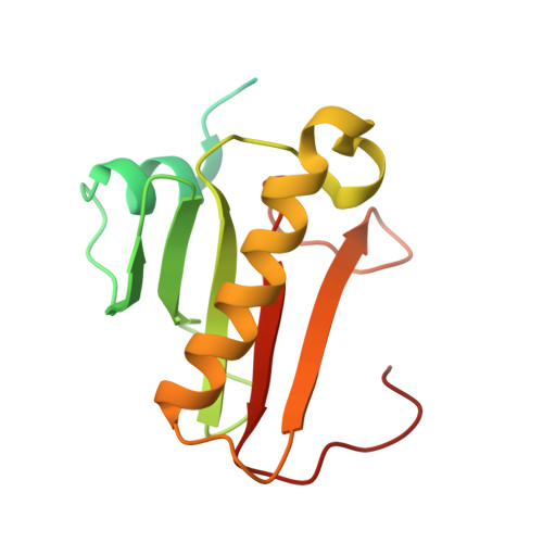
6YPX

Human histidine triad nucleotide-binding protein 2 (hHINT2) refined to 2.11 A in C2221 space group


Experimental Data Snapshot

  • Method: X-RAY DIFFRACTION
  • Resolution: 2.11 Å
  • R-Value Free: 0.184 
  • R-Value Work: 0.148 

Starting Model: experimental
View more details

wwPDB Validation   3D Report Full Report


Ligand Structure Quality Assessment 


This is version 1.2 of the entry. See complete history


Literature

Biochemical, crystallographic and biophysical characterization of histidine triad nucleotide-binding protein 2 with different ligands including a non-hydrolyzable analog of Ap4A.

Dolot, R.Krakowiak, A.Kaczmarek, R.Wlodarczyk, A.Pichlak, M.Nawrot, B.

(2021) Biochim Biophys Acta Gen Subj 1865: 129968-129968

  • DOI: https://doi.org/10.1016/j.bbagen.2021.129968
  • Primary Citation of Related Structures:  
    6YI0, 6YPR, 6YPX, 6YQD, 6YQM, 6YVP

  • PubMed Abstract: 

    Human HINT2 is an important mitochondrial enzyme involved in many processes such as apoptosis and bioenergetics, but its endogenous substrates and the three-dimensional structure of the full-length protein have not been identified yet. An HPLC assay was used to test the hydrolytic activity of HINT2 against various adenosine, guanosine, and 2'-deoxyguanosine derivatives containing phosphate bonds of different types and different leaving groups. Data on binding affinity were obtained by microscale thermophoresis (MST). Crystal structures of HINT2, in its apo form and with a dGMP ligand, were resolved to atomic resolution. HINT2 substrate specificity was similar to that of HINT1, but with the major exception of remarkable discrimination against substrates lacking the 2'-hydroxyl group. The biochemical results were consistent with binding affinity measurements. They showed a similar binding strength of AMP and GMP to HINT2, and much weaker binding of dGMP, in contrast to HINT1. A non-hydrolyzable analog of Ap4A (JB419) interacted with both proteins with similar K d and Ap4A is the signaling molecule that can interact with hHINT1 and regulate the activity of some transcription factors. Several forms of homo- and heterodimers of different lengths of N-terminally truncated polypeptides resulting from degradation of the full-length protein were described. Ser144 in HINT2 appeared to be functionally equivalent to Ser107 in HINT1 by supporting the protonation of the leaving group in the hydrolytic mechanism of HINT2. Our results should be considered in future studies on the natural function of HINT2 and its role in nucleotide prodrug processing.


  • Organizational Affiliation

    Centre of Molecular and Macromolecular Studies, Polish Academy of Sciences, Sienkiewicza 112, 90-363 Lodz, Poland. Electronic address: [email protected].


Macromolecules
Find similar proteins by:  (by identity cutoff)  |  3D Structure
Entity ID: 1
MoleculeChains Sequence LengthOrganismDetailsImage
Histidine triad nucleotide-binding protein 2, mitochondrialA [auth AAA],
B [auth BBB],
C [auth CCC],
D [auth DDD]
163Homo sapiensMutation(s): 0 
Gene Names: HINT2
EC: 3 (PDB Primary Data), 3.9.1 (UniProt)
UniProt & NIH Common Fund Data Resources
Find proteins for Q9BX68 (Homo sapiens)
Explore Q9BX68 
Go to UniProtKB:  Q9BX68
PHAROS:  Q9BX68
GTEx:  ENSG00000137133 
Entity Groups  
Sequence Clusters30% Identity50% Identity70% Identity90% Identity95% Identity100% Identity
UniProt GroupQ9BX68
Sequence Annotations
Expand
  • Reference Sequence
Small Molecules
Ligands 3 Unique
IDChains Name / Formula / InChI Key2D Diagram3D Interactions
CAC (Subject of Investigation/LOI)
Query on CAC

Download Ideal Coordinates CCD File 
E [auth AAA],
I [auth DDD]
CACODYLATE ION
C2 H6 As O2
OGGXGZAMXPVRFZ-UHFFFAOYSA-M
GOL (Subject of Investigation/LOI)
Query on GOL

Download Ideal Coordinates CCD File 
F [auth BBB],
G [auth CCC]
GLYCEROL
C3 H8 O3
PEDCQBHIVMGVHV-UHFFFAOYSA-N
NA (Subject of Investigation/LOI)
Query on NA

Download Ideal Coordinates CCD File 
H [auth CCC],
J [auth DDD]
SODIUM ION
Na
FKNQFGJONOIPTF-UHFFFAOYSA-N
Experimental Data & Validation

Experimental Data

  • Method: X-RAY DIFFRACTION
  • Resolution: 2.11 Å
  • R-Value Free: 0.184 
  • R-Value Work: 0.148 
  • Space Group: C 2 2 21
Unit Cell:
Length ( Å )Angle ( ˚ )
a = 138.411α = 90
b = 153.992β = 90
c = 74.785γ = 90
Software Package:
Software NamePurpose
REFMACrefinement
XDSdata reduction
Aimlessdata scaling
MOLREPphasing

Structure Validation

View Full Validation Report


Ligand Structure Quality Assessment 


Entry History & Funding Information

Deposition Data


Funding OrganizationLocationGrant Number
Ministry of Science and Higher Education (Poland)PolandN N204 516139

Revision History  (Full details and data files)

  • Version 1.0: 2020-04-29
    Type: Initial release
  • Version 1.1: 2021-11-10
    Changes: Data collection, Database references, Derived calculations
  • Version 1.2: 2024-01-24
    Changes: Data collection, Derived calculations, Refinement description