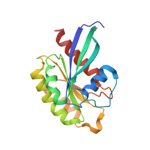
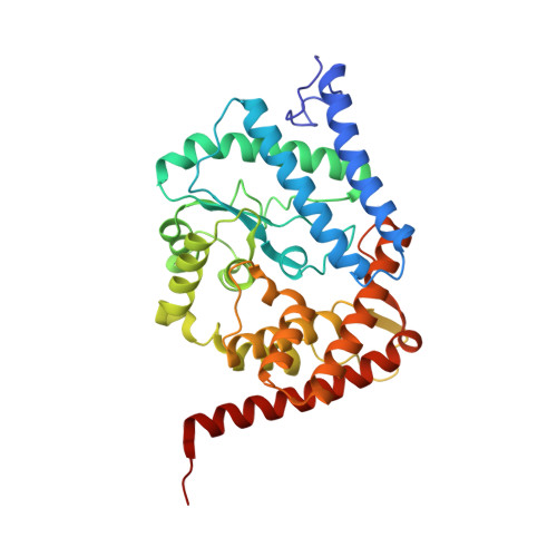
6YX5

Structure of DrrA from Legionella pneumophilia in complex with human Rab8a


Experimental Data Snapshot

  • Method: X-RAY DIFFRACTION
  • Resolution: 2.14 Å
  • R-Value Free: 0.238 
  • R-Value Work: 0.207 
  • R-Value Observed: 0.208 

Starting Models: experimental
View more details

wwPDB Validation   3D Report Full Report


Ligand Structure Quality Assessment 


This is version 1.3 of the entry. See complete history


Literature

Rab1-AMPylation by Legionella DrrA is allosterically activated by Rab1.

Du, J.Wrisberg, M.V.Gulen, B.Stahl, M.Pett, C.Hedberg, C.Lang, K.Schneider, S.Itzen, A.

(2021) Nat Commun 12: 460-460

  • DOI: https://doi.org/10.1038/s41467-020-20702-2
  • Primary Citation of Related Structures:  
    6YX5

  • PubMed Abstract: 

    Legionella pneumophila infects eukaryotic cells by forming a replicative organelle - the Legionella containing vacuole. During this process, the bacterial protein DrrA/SidM is secreted and manipulates the activity and post-translational modification (PTM) states of the vesicular trafficking regulator Rab1. As a result, Rab1 is modified with an adenosine monophosphate (AMP), and this process is referred to as AMPylation. Here, we use a chemical approach to stabilise low-affinity Rab:DrrA complexes in a site-specific manner to gain insight into the molecular basis of the interaction between the Rab protein and the AMPylation domain of DrrA. The crystal structure of the Rab:DrrA complex reveals a previously unknown non-conventional Rab-binding site (NC-RBS). Biochemical characterisation demonstrates allosteric stimulation of the AMPylation activity of DrrA via Rab binding to the NC-RBS. We speculate that allosteric control of DrrA could in principle prevent random and potentially cytotoxic AMPylation in the host, thereby perhaps ensuring efficient infection by Legionella.


  • Organizational Affiliation

    Center for Integrated Protein Science Munich (CIPSM), Department of Chemistry, Technical University of Munich, Garching, 85748, Germany.


Macromolecules
Find similar proteins by:  (by identity cutoff)  |  3D Structure
Entity ID: 1
MoleculeChains Sequence LengthOrganismDetailsImage
Ras-related protein Rab-8A174Homo sapiensMutation(s): 0 
Gene Names: Rab8a
EC: 3.6.5.2
UniProt & NIH Common Fund Data Resources
Find proteins for P61006 (Homo sapiens)
Explore P61006 
Go to UniProtKB:  P61006
PHAROS:  P61006
GTEx:  ENSG00000167461 
Entity Groups  
Sequence Clusters30% Identity50% Identity70% Identity90% Identity95% Identity100% Identity
UniProt GroupP61006
Sequence Annotations
Expand
  • Reference Sequence
Find similar proteins by:  (by identity cutoff)  |  3D Structure
Entity ID: 2
MoleculeChains Sequence LengthOrganismDetailsImage
Multifunctional virulence effector protein DrrA340Legionella pneumophilaMutation(s): 1 
EC: 2.7.7.108
UniProt
Find proteins for Q29ST3 (Legionella pneumophila)
Explore Q29ST3 
Go to UniProtKB:  Q29ST3
Entity Groups  
Sequence Clusters30% Identity50% Identity70% Identity90% Identity95% Identity100% Identity
UniProt GroupQ29ST3
Sequence Annotations
Expand
  • Reference Sequence
Small Molecules
Ligands 5 Unique
IDChains Name / Formula / InChI Key2D Diagram3D Interactions
GNP
Query on GNP

Download Ideal Coordinates CCD File 
D [auth A]PHOSPHOAMINOPHOSPHONIC ACID-GUANYLATE ESTER
C10 H17 N6 O13 P3
UQABYHGXWYXDTK-UUOKFMHZSA-N
LJN
Query on LJN

Download Ideal Coordinates CCD File 
F [auth A][(2~{R},3~{S},4~{R},5~{R})-5-[4-(acetamidomethyl)-1,2,3-triazol-1-yl]-3,4-bis(oxidanyl)oxolan-2-yl]methyl dihydrogen phosphate
C10 H17 N4 O8 P
WKSDLIPBAPRDGN-ZYUZMQFOSA-N
GLC
Query on GLC

Download Ideal Coordinates CCD File 
E [auth A],
I [auth B],
J [auth B]
alpha-D-glucopyranose
C6 H12 O6
WQZGKKKJIJFFOK-DVKNGEFBSA-N
SO4
Query on SO4

Download Ideal Coordinates CCD File 
G [auth A],
H [auth A],
K [auth B],
L [auth B],
M [auth B]
SULFATE ION
O4 S
QAOWNCQODCNURD-UHFFFAOYSA-L
MG
Query on MG

Download Ideal Coordinates CCD File 
C [auth A]MAGNESIUM ION
Mg
JLVVSXFLKOJNIY-UHFFFAOYSA-N
Experimental Data & Validation

Experimental Data

  • Method: X-RAY DIFFRACTION
  • Resolution: 2.14 Å
  • R-Value Free: 0.238 
  • R-Value Work: 0.207 
  • R-Value Observed: 0.208 
  • Space Group: P 3 2 1
Unit Cell:
Length ( Å )Angle ( ˚ )
a = 142.309α = 90
b = 142.309β = 90
c = 76.616γ = 120
Software Package:
Software NamePurpose
XDSdata reduction
REFMACrefinement
PDB_EXTRACTdata extraction
XDSdata scaling
PHASERphasing

Structure Validation

View Full Validation Report


Ligand Structure Quality Assessment 


Entry History & Funding Information

Deposition Data


Funding OrganizationLocationGrant Number
German Research Foundation (DFG)GermanySFB1035
German Research Foundation (DFG)GermanyEXC114

Revision History  (Full details and data files)

  • Version 1.0: 2020-12-23
    Type: Initial release
  • Version 1.1: 2021-02-03
    Changes: Database references
  • Version 1.2: 2024-01-24
    Changes: Data collection, Database references, Refinement description
  • Version 1.3: 2024-11-20
    Changes: Structure summary