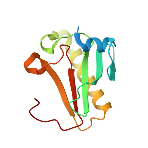
6J65

Crystal structure of human HINT1 mutant complexing with AP4A II


Experimental Data Snapshot

  • Method: X-RAY DIFFRACTION
  • Resolution: 1.42 Å
  • R-Value Free: 0.206 
  • R-Value Work: 0.180 
  • R-Value Observed: 0.181 

Starting Model: experimental
View more details

wwPDB Validation   3D Report Full Report


Ligand Structure Quality Assessment 


This is version 1.2 of the entry. See complete history


Literature

Second messenger Ap4A polymerizes target protein HINT1 to transduce signals in Fc epsilon RI-activated mast cells.

Yu, J.Liu, Z.Liang, Y.Luo, F.Zhang, J.Tian, C.Motzik, A.Zheng, M.Kang, J.Zhong, G.Liu, C.Fang, P.Guo, M.Razin, E.Wang, J.

(2019) Nat Commun 10: 4664-4664

  • DOI: https://doi.org/10.1038/s41467-019-12710-8
  • Primary Citation of Related Structures:  
    5ED3, 5ED6, 6J53, 6J58, 6J5S, 6J5Z, 6J64, 6J65

  • PubMed Abstract: 

    Signal transduction systems enable organisms to monitor their external environments and accordingly adjust the cellular processes. In mast cells, the second messenger Ap 4 A binds to the histidine triad nucleotide-binding protein 1 (HINT1), disrupts its interaction with the microphthalmia-associated transcription factor (MITF), and eventually activates the transcription of genes downstream of MITF in response to immunostimulation. How the HINT1 protein recognizes and is regulated by Ap 4 A remain unclear. Here, using eight crystal structures, biochemical experiments, negative stain electron microscopy, and cellular experiments, we report that Ap 4 A specifically polymerizes HINT1 in solution and in activated rat basophilic leukemia cells. The polymerization interface overlaps with the area on HINT1 for MITF interaction, suggesting a possible competitive mechanism to release MITF for transcriptional activation. The mechanism depends precisely on the length of the phosphodiester linkage of Ap 4 A. These results highlight a direct polymerization signaling mechanism by the second messenger.


  • Organizational Affiliation

    State Key Laboratory of Bioorganic and Natural Products Chemistry, Center for Excellence in Molecular Synthesis, Shanghai Institute of Organic Chemistry, University of Chinese Academy of Sciences, Chinese Academy of Sciences, 345 Lingling Road, Shanghai, 200032, China.


Macromolecules
Find similar proteins by:  (by identity cutoff)  |  3D Structure
Entity ID: 1
MoleculeChains Sequence LengthOrganismDetailsImage
Histidine triad nucleotide-binding protein 1A,
B,
C [auth D],
D [auth E]
128Homo sapiensMutation(s): 1 
Gene Names: HINT1HINTPKCI1PRKCNH1
EC: 3 (PDB Primary Data), 3.4.22 (UniProt), 3.9.1 (UniProt)
UniProt & NIH Common Fund Data Resources
Find proteins for P49773 (Homo sapiens)
Explore P49773 
Go to UniProtKB:  P49773
PHAROS:  P49773
GTEx:  ENSG00000169567 
Entity Groups  
Sequence Clusters30% Identity50% Identity70% Identity90% Identity95% Identity100% Identity
UniProt GroupP49773
Sequence Annotations
Expand
  • Reference Sequence
Small Molecules
Ligands 2 Unique
IDChains Name / Formula / InChI Key2D Diagram3D Interactions
B4P
Query on B4P

Download Ideal Coordinates CCD File 
E [auth B]BIS(ADENOSINE)-5'-TETRAPHOSPHATE
C20 H28 N10 O19 P4
YOAHKNVSNCMZGQ-XPWFQUROSA-N
EPE
Query on EPE

Download Ideal Coordinates CCD File 
F [auth E]4-(2-HYDROXYETHYL)-1-PIPERAZINE ETHANESULFONIC ACID
C8 H18 N2 O4 S
JKMHFZQWWAIEOD-UHFFFAOYSA-N
Experimental Data & Validation

Experimental Data

  • Method: X-RAY DIFFRACTION
  • Resolution: 1.42 Å
  • R-Value Free: 0.206 
  • R-Value Work: 0.180 
  • R-Value Observed: 0.181 
  • Space Group: P 1
Unit Cell:
Length ( Å )Angle ( ˚ )
a = 46.3α = 98.06
b = 46.573β = 89.82
c = 64.246γ = 118.25
Software Package:
Software NamePurpose
PHENIXrefinement
HKL-2000data scaling
PDB_EXTRACTdata extraction
HKL-2000data reduction

Structure Validation

View Full Validation Report


Ligand Structure Quality Assessment 


Entry History & Funding Information

Deposition Data


Funding OrganizationLocationGrant Number
National Natural Science Foundation of ChinaChina21778067
National Natural Science Foundation of ChinaChina21778064

Revision History  (Full details and data files)

  • Version 1.0: 2019-09-25
    Type: Initial release
  • Version 1.1: 2020-04-08
    Changes: Database references
  • Version 1.2: 2023-11-22
    Changes: Data collection, Database references, Refinement description