Targeting a Cryptic Pocket in a Protein-Protein Contact by Disulfide-Induced Rupture of a Homodimeric Interface.
Nguyen, D., Xie, X., Jakobi, S., Terwesten, F., Metz, A., Nguyen, T.X.P., Palchykov, V.A., Heine, A., Reuter, K., Klebe, G.(2021) ACS Chem Biol 16: 1090-1098
- PubMed: 34081441
- DOI: https://doi.org/10.1021/acschembio.1c00296
- Primary Citation of Related Structures:
4JBR, 7A0B, 7A3V, 7A3X, 7A4K, 7A4X, 7A6D, 7A9E, 7ADN - PubMed Abstract:
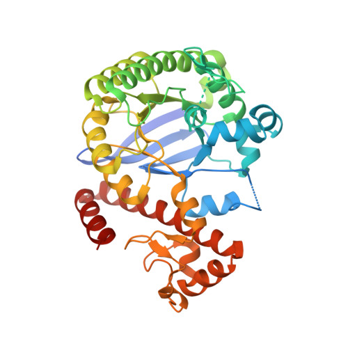
Interference with protein-protein interfaces represents an attractive as well as challenging option for therapeutic intervention and drug design. The enzyme tRNA-guanine transglycosylase, a target to fight Shigellosis, is only functional as a homodimer. Although we previously produced monomeric variants by site-directed mutagenesis, we only crystallized the functional dimer, simply because upon crystallization the local protein concentration increases and favors formation of the dimer interface, which represents an optimal and highly stable packing of the protein in the solid state. Unfortunately, this prevents access to structural information about the interface geometry in its monomeric state and complicates the development of modulators that can interfere with and prevent dimer formation. Here, we report on a cysteine-containing protein variant in which, under oxidizing conditions, a disulfide linkage is formed. This reinforces a novel packing geometry of the enzyme. In this captured quasi-monomeric state, the monomer units arrange in a completely different way and, thus, expose a loop-helix motif, originally embedded into the old interface, now to the surface. The motif adopts a geometry incompatible with the original dimer formation. Via the soaking of fragments into the crystals, we identified several hits accommodating a cryptic binding site next to the loop-helix motif and modulated its structural features. Our study demonstrates the druggability of the interface by breaking up the homodimeric protein using an introduced disulfide cross-link. By rational concepts, we increased the potency of these fragments to a level where we confirmed their binding by NMR to a nondisulfide-linked TGT variant. The idea of intermediately introducing a disulfide linkage may serve as a general concept of how to transform a homodimer interface into a quasi-monomeric state and give access to essential structural and design information.
Organizational Affiliation:
Institut für Pharmazeutische Chemie, Philipps-Universität Marburg, Marbacher Weg 8, 35032 Marburg, Germany.


