7ACD

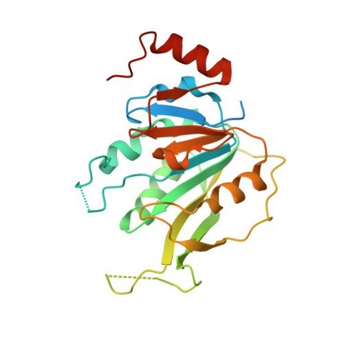
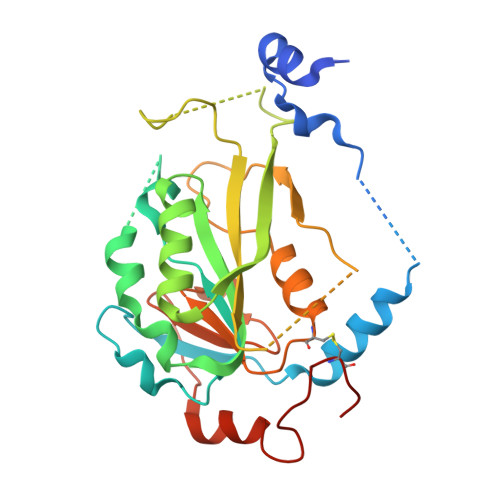
Crystal structure of the human METTL3-METTL14 complex with compound T30 (UZH1a)


Experimental Data Snapshot

  • Method: X-RAY DIFFRACTION
  • Resolution: 2.50 Å
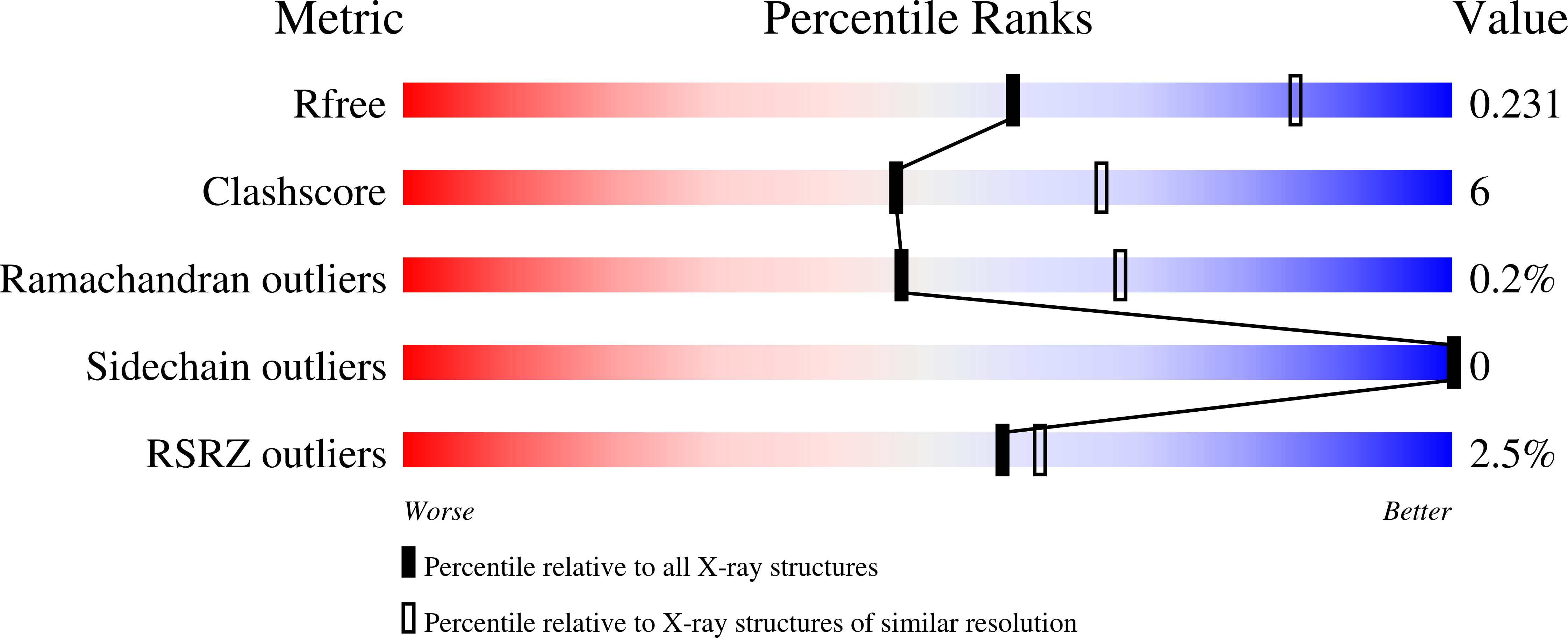
  • R-Value Free: 0.232 
  • R-Value Work: 0.187 
  • R-Value Observed: 0.190 

Starting Model: experimental
View more details

wwPDB Validation   3D Report Full Report


Ligand Structure Quality Assessment 


This is version 2.3 of the entry. See complete history


Literature

METTL3 Inhibitors for Epitranscriptomic Modulation of Cellular Processes.

Moroz-Omori, E.V.Huang, D.Kumar Bedi, R.Cheriyamkunnel, S.J.Bochenkova, E.Dolbois, A.Rzeczkowski, M.D.Li, Y.Wiedmer, L.Caflisch, A.

(2021) ChemMedChem 16: 3035-3043

  • DOI: https://doi.org/10.1002/cmdc.202100291
  • Primary Citation of Related Structures:  
    7ACD

  • PubMed Abstract: 

    The methylase METTL3 is the writer enzyme of the N 6 -methyladenosine (m 6 A) modification of RNA. Using a structure-based drug discovery approach, we identified a METTL3 inhibitor with potency in a biochemical assay of 280 nM, while its enantiomer is 100 times less active. We observed a dose-dependent reduction in the m 6 A methylation level of mRNA in several cell lines treated with the inhibitor already after 16 h of treatment, which lasted for at least 6 days. Importantly, the prolonged incubation (up to 6 days) with the METTL3 inhibitor did not alter levels of other RNA modifications (i. e., m 1 A, m 6 A m , m 7 G), suggesting selectivity of the developed compound towards other RNA methyltransferases.


  • Organizational Affiliation

    Department of Biochemistry, University of Zurich, Winterthurerstrasse 190, 8057, Zurich, Switzerland.


Macromolecules
Find similar proteins by:  (by identity cutoff)  |  3D Structure
Entity ID: 1
MoleculeChains Sequence LengthOrganismDetailsImage
N6-adenosine-methyltransferase catalytic subunit246Homo sapiensMutation(s): 0 
Gene Names: METTL3MTA70
EC: 2.1.1.348
UniProt & NIH Common Fund Data Resources
Find proteins for Q86U44 (Homo sapiens)
Explore Q86U44 
Go to UniProtKB:  Q86U44
PHAROS:  Q86U44
GTEx:  ENSG00000165819 
Entity Groups  
Sequence Clusters30% Identity50% Identity70% Identity90% Identity95% Identity100% Identity
UniProt GroupQ86U44
Sequence Annotations
Expand
  • Reference Sequence
Find similar proteins by:  (by identity cutoff)  |  3D Structure
Entity ID: 2
MoleculeChains Sequence LengthOrganismDetailsImage
N6-adenosine-methyltransferase non-catalytic subunit290Homo sapiensMutation(s): 0 
Gene Names: METTL14KIAA1627
UniProt & NIH Common Fund Data Resources
Find proteins for Q9HCE5 (Homo sapiens)
Explore Q9HCE5 
Go to UniProtKB:  Q9HCE5
PHAROS:  Q9HCE5
GTEx:  ENSG00000145388 
Entity Groups  
Sequence Clusters30% Identity50% Identity70% Identity90% Identity95% Identity100% Identity
UniProt GroupQ9HCE5
Sequence Annotations
Expand
  • Reference Sequence
Small Molecules
Ligands 3 Unique
IDChains Name / Formula / InChI Key2D Diagram3D Interactions
R72 (Subject of Investigation/LOI)
Query on R72

Download Ideal Coordinates CCD File 
C [auth A]4-[(4,4-dimethylpiperidin-1-yl)methyl]-2-oxidanyl-~{N}-[[(3~{R})-3-oxidanyl-1-[6-[(phenylmethyl)amino]pyrimidin-4-yl]piperidin-3-yl]methyl]benzamide
C32 H42 N6 O3
PWYRDVXYAOGDNK-JGCGQSQUSA-N
ACT
Query on ACT

Download Ideal Coordinates CCD File 
E [auth B]ACETATE ION
C2 H3 O2
QTBSBXVTEAMEQO-UHFFFAOYSA-M
MG
Query on MG

Download Ideal Coordinates CCD File 
D [auth B]MAGNESIUM ION
Mg
JLVVSXFLKOJNIY-UHFFFAOYSA-N
Experimental Data & Validation

Experimental Data

  • Method: X-RAY DIFFRACTION
  • Resolution: 2.50 Å
  • R-Value Free: 0.232 
  • R-Value Work: 0.187 
  • R-Value Observed: 0.190 
  • Space Group: P 32 2 1
Unit Cell:
Length ( Å )Angle ( ˚ )
a = 63.8α = 90
b = 63.8β = 90
c = 224.92γ = 120
Software Package:
Software NamePurpose
XDSdata reduction
XSCALEdata scaling
PHASERphasing
PHENIXrefinement

Structure Validation

View Full Validation Report


Ligand Structure Quality Assessment 


Entry History & Funding Information

Deposition Data


Funding OrganizationLocationGrant Number
Swiss National Science FoundationSwitzerland310030B_189363

Revision History  (Full details and data files)

  • Version 1.0: 2020-10-28
    Type: Initial release
  • Version 1.1: 2020-11-04
    Changes: Database references
  • Version 1.2: 2020-11-11
    Changes: Database references
  • Version 2.0: 2021-03-24
    Type: Coordinate replacement
    Reason: Ligand geometry
    Changes: Advisory, Atomic model, Data collection, Database references, Derived calculations, Refinement description, Structure summary
  • Version 2.1: 2021-07-21
    Changes: Database references
  • Version 2.2: 2021-10-13
    Changes: Data collection, Database references
  • Version 2.3: 2024-01-31
    Changes: Data collection, Database references, Refinement description