7CC8

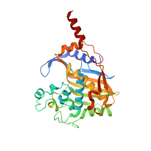
Crystal structure of White Spot Syndrome Virus Thymidylate Synthase - Apo form


Experimental Data Snapshot

  • Method: X-RAY DIFFRACTION
  • Resolution: 2.30 Å
  • R-Value Free: 0.209 
  • R-Value Work: 0.185 

Starting Model: experimental
View more details

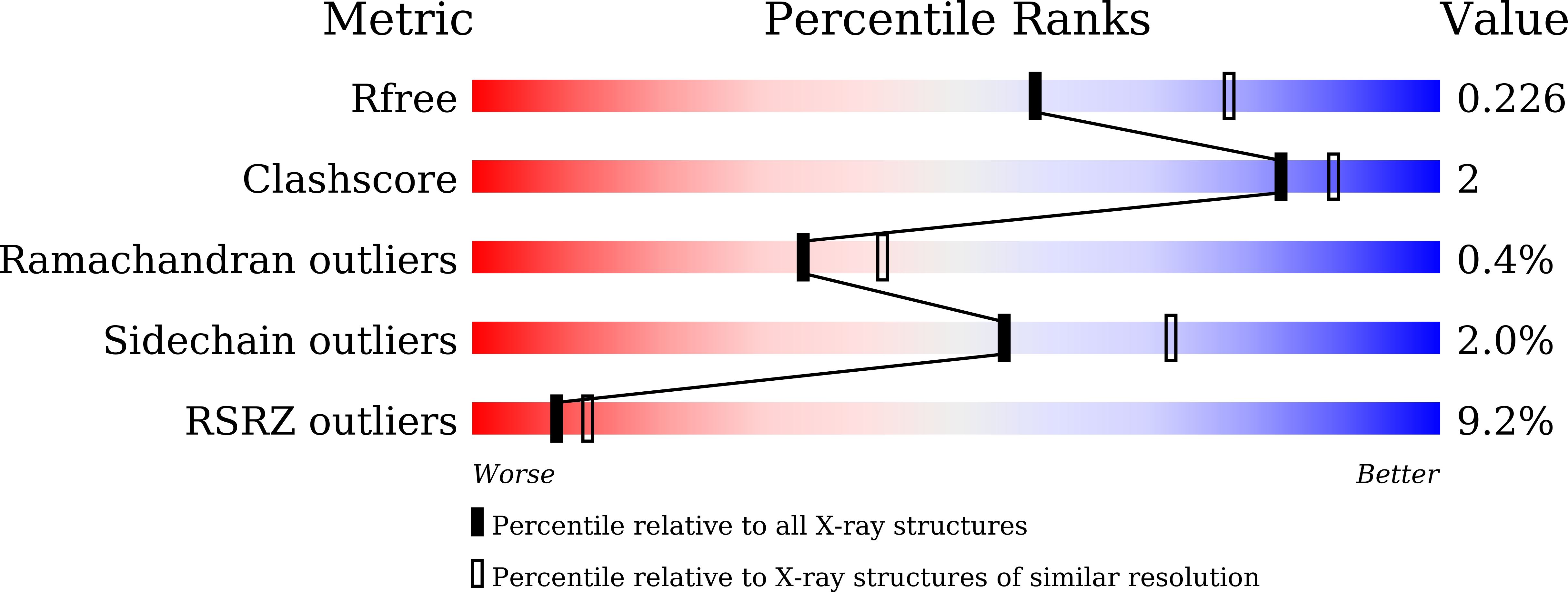
wwPDB Validation   3D Report Full Report


This is version 1.2 of the entry. See complete history


Literature

Structure analysis of thymidylate synthase from white spot syndrome virus reveals WSSV-specific structural elements.

Panchal, V.Kumar, S.Hossain, S.N.Vasudevan, D.

(2021) Int J Biol Macromol 167: 1168-1175

  • DOI: https://doi.org/10.1016/j.ijbiomac.2020.11.071
  • Primary Citation of Related Structures:  
    7CC8, 7CCA

  • PubMed Abstract: 

    White spot syndrome virus (WSSV), the causative agent of white spot disease (WSD) severely affecting crustacean life forms, is highly contagious and forms the principal cause of massive economic losses in the shrimp aquaculture industry. Previous studies have demonstrated thymidylate synthase as a successful anti-cancer therapeutic drug target, leading to various anti-cancer drugs. The differential utilization of nucleotide precursors between white spot syndrome virus and shrimp encouraged us to analyze WSSV-thymidylate synthase (wTS). Here, we report the crystal structures of wTS in its apo-form and as a ternary complex with deoxyuridine monophosphate (dUMP) and methotrexate at a resolution of 2.35 Å and 2.6 Å, respectively. wTS possesses a fold characteristic to known thymidylate synthase (TS) structures. Like other TS structures, the apo-form of wTS displays an open conformation, whereas the wTS ternary complex attains a closed conformation. While the C-terminal loop maintains a typical distance from methotrexate, the Sγ atom of the catalytic Cys is positioned farther from the C6 atom of dUMP. Altogether, we report the first TS structure from a crustacean virus and highlight its distinction from shrimp and other TS structures.


  • Organizational Affiliation

    Institute of Life Sciences, Bhubaneswar 751023, Odisha, India.


Macromolecules
Find similar proteins by:  (by identity cutoff)  |  3D Structure
Entity ID: 1
MoleculeChains Sequence LengthOrganismDetailsImage
Thymidylate Synthase
A, B
297Shrimp white spot syndrome virus (isolate Tongan)Mutation(s): 0 
EC: 2.1.1.45
UniProt
Find proteins for Q77J90 (White spot syndrome virus (isolate Shrimp/China/Tongan/1996))
Explore Q77J90 
Go to UniProtKB:  Q77J90
Entity Groups  
Sequence Clusters30% Identity50% Identity70% Identity90% Identity95% Identity100% Identity
UniProt GroupQ77J90
Sequence Annotations
Expand
  • Reference Sequence
Small Molecules
Ligands 2 Unique
IDChains Name / Formula / InChI Key2D Diagram3D Interactions
PGE
Query on PGE

Download Ideal Coordinates CCD File 
H [auth A],
J [auth B],
K [auth B]
TRIETHYLENE GLYCOL
C6 H14 O4
ZIBGPFATKBEMQZ-UHFFFAOYSA-N
SO4
Query on SO4

Download Ideal Coordinates CCD File 
C [auth A]
D [auth A]
E [auth A]
F [auth A]
G [auth A]
C [auth A],
D [auth A],
E [auth A],
F [auth A],
G [auth A],
I [auth B]
SULFATE ION
O4 S
QAOWNCQODCNURD-UHFFFAOYSA-L
Experimental Data & Validation

Experimental Data

  • Method: X-RAY DIFFRACTION
  • Resolution: 2.30 Å
  • R-Value Free: 0.209 
  • R-Value Work: 0.185 
  • Space Group: P 43 21 2
Unit Cell:
Length ( Å )Angle ( ˚ )
a = 91.801α = 90
b = 91.801β = 90
c = 233.578γ = 90
Software Package:
Software NamePurpose
REFMACrefinement
PDB_EXTRACTdata extraction
iMOSFLMdata reduction
Aimlessdata scaling
MOLREPphasing

Structure Validation

View Full Validation Report


Entry History & Funding Information

Deposition Data


Funding OrganizationLocationGrant Number
Department of Biotechnology (DBT, India)IndiaBT/PR23375/AAQ/3/849/2017

Revision History  (Full details and data files)

  • Version 1.0: 2020-11-25
    Type: Initial release
  • Version 1.1: 2021-01-27
    Changes: Database references
  • Version 1.2: 2023-11-29
    Changes: Data collection, Database references, Refinement description