7CL3

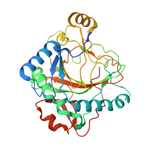
The crystal structure of KanJ in complex with kanamycin B


Experimental Data Snapshot

  • Method: X-RAY DIFFRACTION
  • Resolution: 2.20 Å
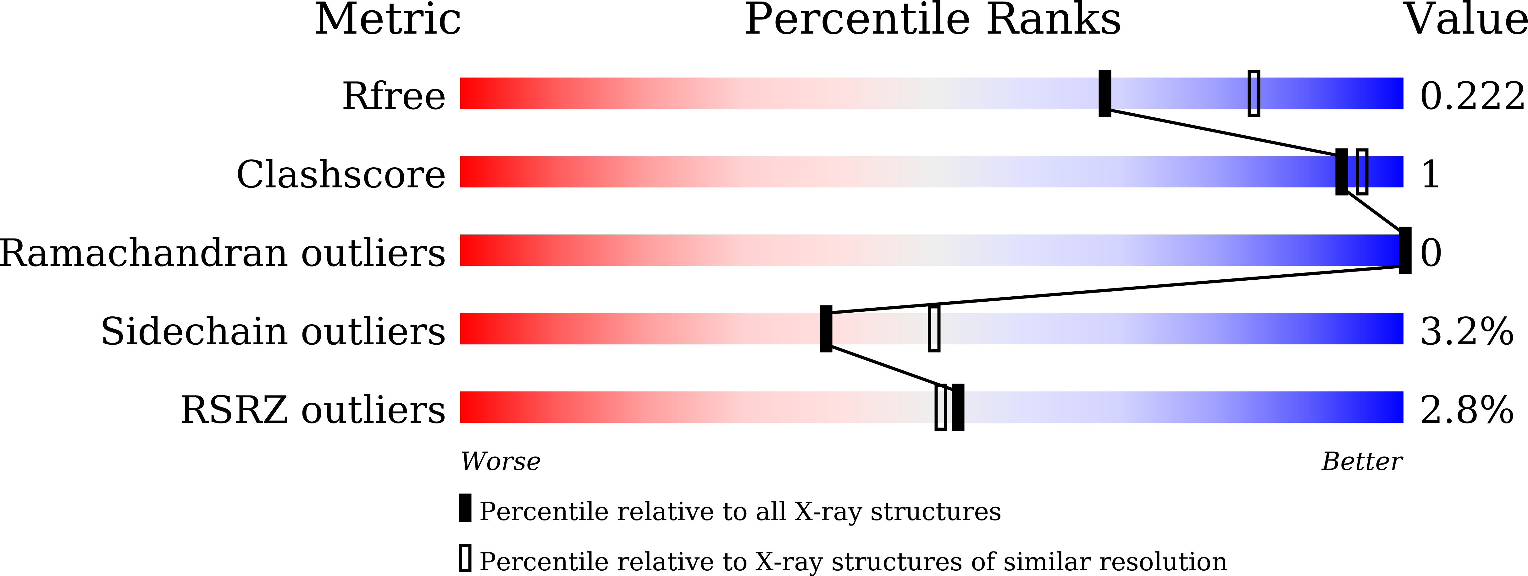
  • R-Value Free: 0.217 
  • R-Value Work: 0.194 
  • R-Value Observed: 0.196 

Starting Model: experimental
View more details

wwPDB Validation   3D Report Full Report


Ligand Structure Quality Assessment 


This is version 1.3 of the entry. See complete history


Literature

Stepwise Post-glycosylation Modification of Sugar Moieties in Kanamycin Biosynthesis.

Kudo, F.Kitayama, Y.Miyanaga, A.Numakura, M.Eguchi, T.

(2021) Chembiochem 22: 1668-1675

  • DOI: https://doi.org/10.1002/cbic.202000839
  • Primary Citation of Related Structures:  
    7CL2, 7CL3, 7CL4, 7CL5, 7CL6

  • PubMed Abstract: 

    Kanamycin A is the major 2-deoxystreptamine (2DOS)-containing aminoglycoside antibiotic produced by Streptomyces kanamyceticus. The 2DOS moiety is linked with 6-amino-6-deoxy-d-glucose (6ADG) at O-4 and 3-amino-3-deoxy-d-glucose at O-6. Because the 6ADG moiety is derived from d-glucosamine (GlcN), deamination at C-2 and introduction of C-6-NH 2 are required in the biosynthesis. A dehydrogenase, KanQ, and an aminotransferase, KanB, are presumed to be responsible for the introduction of C-6-NH 2 , although the substrates have not been identified. Here, we examined the substrate specificity of KanQ to better understand the biosynthetic pathway. It was found that KanQ oxidized kanamycin C more efficiently than the 3''-deamino derivative. Furthermore, the substrate specificity of an oxygenase, KanJ, that is responsible for deamination at C-2 of the GlcN moiety was examined, and the crystal structure of KanJ was determined. It was found that C-6-NH 2 is important for substrate recognition by KanJ. Thus, the modification of the GlcN moiety occurs after pseudo-trisaccharide formation, followed by the introduction of C-6-NH 2 by KanQ/KanB and deamination at C-2 by KanJ.


  • Organizational Affiliation

    Department of Chemistry, Tokyo Institute of Technology, 2-12-1 O-okayama, Meguro-ku, Tokyo, 152-8551, Japan.


Macromolecules
Find similar proteins by:  (by identity cutoff)  |  3D Structure
Entity ID: 1
MoleculeChains Sequence LengthOrganismDetailsImage
Kanamycin B dioxygenase
A, B, C, D, E
A, B, C, D, E, F
301Streptomyces kanamyceticusMutation(s): 0 
Gene Names: kanJkacB
EC: 1.14.11.37
UniProt
Find proteins for Q6L732 (Streptomyces kanamyceticus)
Explore Q6L732 
Go to UniProtKB:  Q6L732
Entity Groups  
Sequence Clusters30% Identity50% Identity70% Identity90% Identity95% Identity100% Identity
UniProt GroupQ6L732
Sequence Annotations
Expand
  • Reference Sequence
Small Molecules
Ligands 3 Unique
IDChains Name / Formula / InChI Key2D Diagram3D Interactions
9CS (Subject of Investigation/LOI)
Query on 9CS

Download Ideal Coordinates CCD File 
I [auth A]
L [auth B]
O [auth C]
R [auth D]
U [auth E]
I [auth A],
L [auth B],
O [auth C],
R [auth D],
U [auth E],
X [auth F]
(1R,2S,3S,4R,6S)-4,6-DIAMINO-3-[(3-AMINO-3-DEOXY-ALPHA-D-GLUCOPYRANOSYL)OXY]-2-HYDROXYCYCLOHEXYL 2,6-DIAMINO-2,6-DIDEOXY-ALPHA-D-GLUCOPYRANOSIDE
C18 H37 N5 O10
SKKLOUVUUNMCJE-FQSMHNGLSA-N
SO4 (Subject of Investigation/LOI)
Query on SO4

Download Ideal Coordinates CCD File 
H [auth A]
K [auth B]
N [auth C]
Q [auth D]
T [auth E]
H [auth A],
K [auth B],
N [auth C],
Q [auth D],
T [auth E],
W [auth F]
SULFATE ION
O4 S
QAOWNCQODCNURD-UHFFFAOYSA-L
NI (Subject of Investigation/LOI)
Query on NI

Download Ideal Coordinates CCD File 
G [auth A]
J [auth B]
M [auth C]
P [auth D]
S [auth E]
G [auth A],
J [auth B],
M [auth C],
P [auth D],
S [auth E],
V [auth F]
NICKEL (II) ION
Ni
VEQPNABPJHWNSG-UHFFFAOYSA-N
Experimental Data & Validation

Experimental Data

  • Method: X-RAY DIFFRACTION
  • Resolution: 2.20 Å
  • R-Value Free: 0.217 
  • R-Value Work: 0.194 
  • R-Value Observed: 0.196 
  • Space Group: P 1 21 1
Unit Cell:
Length ( Å )Angle ( ˚ )
a = 50.289α = 90
b = 184.514β = 93.99
c = 109.543γ = 90
Software Package:
Software NamePurpose
REFMACrefinement
PDB_EXTRACTdata extraction
XDSdata reduction
SCALAdata scaling
MOLREPphasing

Structure Validation

View Full Validation Report


Ligand Structure Quality Assessment 


Entry History 

Deposition Data

Revision History  (Full details and data files)

  • Version 1.0: 2021-01-13
    Type: Initial release
  • Version 1.1: 2021-01-20
    Changes: Database references
  • Version 1.2: 2021-05-12
    Changes: Database references
  • Version 1.3: 2023-11-29
    Changes: Data collection, Database references, Refinement description