7CRI

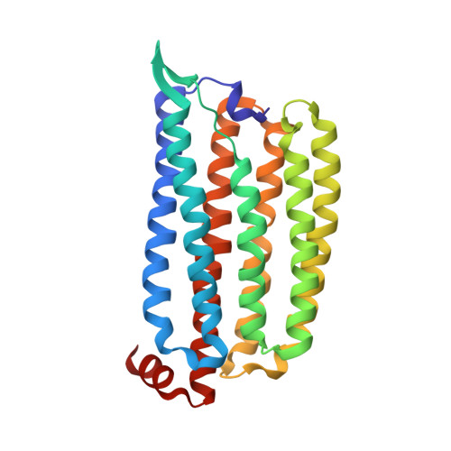
1 ps Structure of Chloride ion pumping rhodopsin (ClR) with NTQ motif


Experimental Data Snapshot

  • Method: X-RAY DIFFRACTION
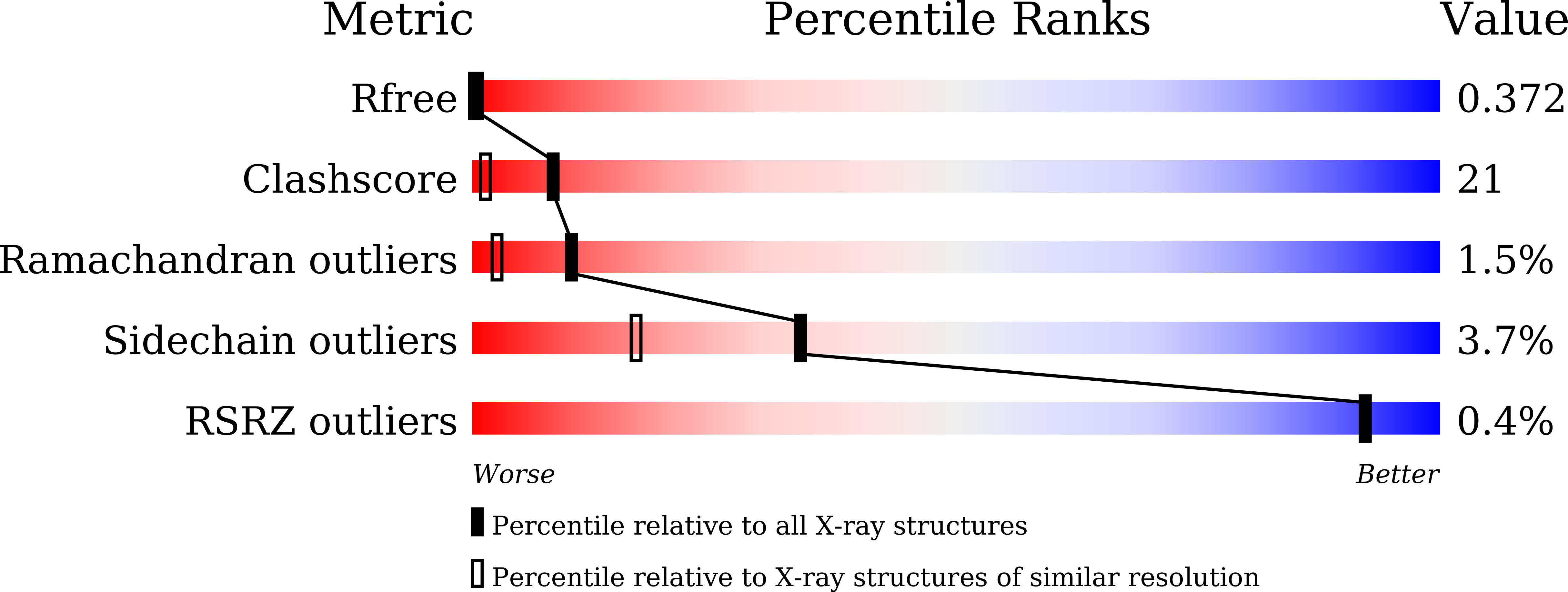
  • Resolution: 1.85 Å
  • R-Value Free: 0.372 
  • R-Value Work: 0.295 
  • R-Value Observed: 0.299 

Starting Model: experimental
View more details

wwPDB Validation   3D Report Full Report


Ligand Structure Quality Assessment 


This is version 1.3 of the entry. See complete history


Literature

Early-stage dynamics of chloride ion-pumping rhodopsin revealed by a femtosecond X-ray laser.

Yun, J.H.Li, X.Yue, J.Park, J.H.Jin, Z.Li, C.Hu, H.Shi, Y.Pandey, S.Carbajo, S.Boutet, S.Hunter, M.S.Liang, M.Sierra, R.G.Lane, T.J.Zhou, L.Weierstall, U.Zatsepin, N.A.Ohki, M.Tame, J.R.H.Park, S.Y.Spence, J.C.H.Zhang, W.Schmidt, M.Lee, W.Liu, H.

(2021) Proc Natl Acad Sci U S A 118

  • DOI: https://doi.org/10.1073/pnas.2020486118
  • Primary Citation of Related Structures:  
    7CRI, 7CRJ, 7CRK, 7CRL, 7CRS, 7CRT, 7CRX, 7CRY

  • PubMed Abstract: 

    Chloride ion-pumping rhodopsin (ClR) in some marine bacteria utilizes light energy to actively transport Cl - into cells. How the ClR initiates the transport is elusive. Here, we show the dynamics of ion transport observed with time-resolved serial femtosecond (fs) crystallography using the Linac Coherent Light Source. X-ray pulses captured structural changes in ClR upon flash illumination with a 550 nm fs-pumping laser. High-resolution structures for five time points (dark to 100 ps after flashing) reveal complex and coordinated dynamics comprising retinal isomerization, water molecule rearrangement, and conformational changes of various residues. Combining data from time-resolved spectroscopy experiments and molecular dynamics simulations, this study reveals that the chloride ion close to the Schiff base undergoes a dissociation-diffusion process upon light-triggered retinal isomerization.


  • Organizational Affiliation

    Department of Biochemistry, College of Life Sciences and Biotechnology, Yonsei University, Seodaemun-gu, 120-749 Seoul, South Korea.


Macromolecules
Find similar proteins by:  (by identity cutoff)  |  3D Structure
Entity ID: 1
MoleculeChains Sequence LengthOrganismDetailsImage
Chloride pumping rhodopsin264Nonlabens marinus S1-08Mutation(s): 0 
Gene Names: ClRNMS_1267
Membrane Entity: Yes 
UniProt
Find proteins for W8VZW3 (Nonlabens marinus S1-08)
Explore W8VZW3 
Go to UniProtKB:  W8VZW3
Entity Groups  
Sequence Clusters30% Identity50% Identity70% Identity90% Identity95% Identity100% Identity
UniProt GroupW8VZW3
Sequence Annotations
Expand
  • Reference Sequence
Small Molecules
Ligands 3 Unique
IDChains Name / Formula / InChI Key2D Diagram3D Interactions
RET
Query on RET

Download Ideal Coordinates CCD File 
D [auth A]RETINAL
C20 H28 O
NCYCYZXNIZJOKI-OVSJKPMPSA-N
OLA
Query on OLA

Download Ideal Coordinates CCD File 
E [auth A]
F [auth A]
G [auth A]
H [auth A]
I [auth A]
E [auth A],
F [auth A],
G [auth A],
H [auth A],
I [auth A],
J [auth A],
K [auth A]
OLEIC ACID
C18 H34 O2
ZQPPMHVWECSIRJ-KTKRTIGZSA-N
CL
Query on CL

Download Ideal Coordinates CCD File 
B [auth A],
C [auth A]
CHLORIDE ION
Cl
VEXZGXHMUGYJMC-UHFFFAOYSA-M
Experimental Data & Validation

Experimental Data

  • Method: X-RAY DIFFRACTION
  • Resolution: 1.85 Å
  • R-Value Free: 0.372 
  • R-Value Work: 0.295 
  • R-Value Observed: 0.299 
  • Space Group: C 1 2 1
Unit Cell:
Length ( Å )Angle ( ˚ )
a = 103.37α = 90
b = 50.09β = 109.69
c = 69.39γ = 90
Software Package:
Software NamePurpose
PHENIXrefinement
CrystFELdata reduction
CrystFELdata scaling
PHENIXphasing

Structure Validation

View Full Validation Report


Ligand Structure Quality Assessment 


Entry History & Funding Information

Deposition Data


Funding OrganizationLocationGrant Number
National Natural Science Foundation of China (NSFC)China11575021,U1930402,21773012
National Research Foundation (NRF, Korea)Korea, Republic Of2019M3E5D6063903, 2017M3A9F6029753, 2018K2A9A2A06024227,2016R1A6A3A04010213
National Science Foundation (NSF, United States)United States1231306,1565180,2030466,DE-AC02-76SF00515

Revision History  (Full details and data files)

  • Version 1.0: 2020-09-30
    Type: Initial release
  • Version 1.1: 2021-04-14
    Changes: Database references
  • Version 1.2: 2023-11-29
    Changes: Advisory, Data collection, Database references, Refinement description
  • Version 1.3: 2024-11-06
    Changes: Structure summary