7EKV

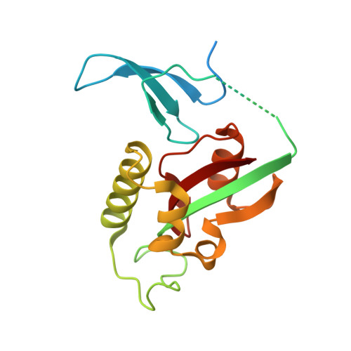
Crystal Structure of human Pin1 complexed with a covalent inhibitor


Experimental Data Snapshot

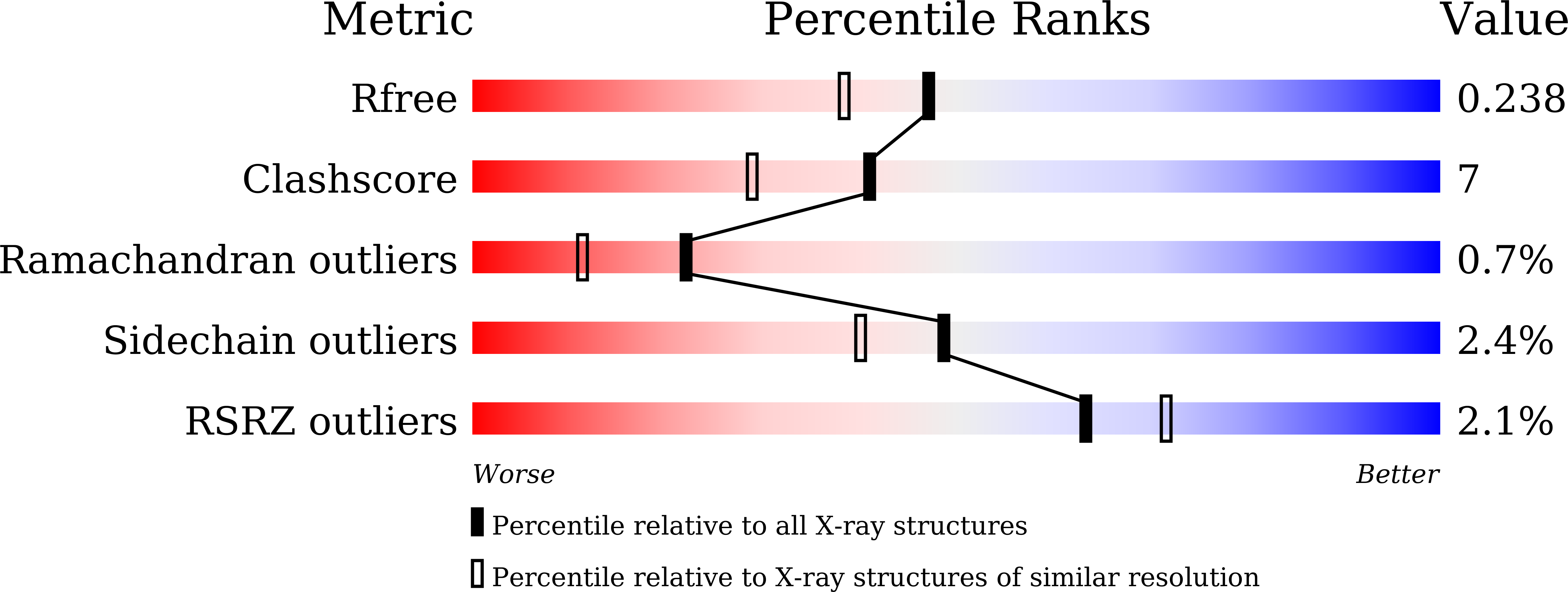
  • Method: X-RAY DIFFRACTION
  • Resolution: 1.95 Å
  • R-Value Free: 0.235 
  • R-Value Work: 0.197 
  • R-Value Observed: 0.201 

Starting Model: experimental
View more details

wwPDB Validation   3D Report Full Report


Ligand Structure Quality Assessment 


This is version 1.3 of the entry. See complete history


Literature

Computational and Structure-Based Development of High Potent Cell-Active Covalent Inhibitor Targeting the Peptidyl-Prolyl Isomerase NIMA-Interacting-1 (Pin1).

Liu, L.Zhu, R.Li, J.Pei, Y.Wang, S.Xu, P.Wang, M.Wen, Y.Zhang, H.Du, D.Ding, H.Jiang, H.Chen, K.Zhou, B.Yu, L.Luo, C.

(2022) J Med Chem 65: 2174-2190

  • DOI: https://doi.org/10.1021/acs.jmedchem.1c01686
  • Primary Citation of Related Structures:  
    7EFJ, 7EFX, 7EKV, 7F0M

  • PubMed Abstract: 

    The unique proline isomerase peptidyl-prolyl isomerase NIMA-interacting-1 (Pin1) is reported to activate numerous cancer-driving pathways simultaneously, and aberrant Pin1 activation is present in many human cancers. Here, we identified a novel hit compound, ZL-Pin01 , that covalently modified Pin1 at Cys113 with an half-maximal inhibitory concentration (IC 50 ) of 1.33 ± 0.07 μM through screening an in-house library. Crystallographic study drove the process of structure-guided optimization and led to the potent inhibitor ZL-Pin13 with an IC 50 of 0.067 ± 0.03 μM. We obtained four co-crystal structures of Pin1 complexed with inhibitors that elucidated the detailed binding mode of the derivatives with Pin1. Interestingly, the co-crystal of Pin1 with ZL-Pin13 obtained by co-crystallization revealed the conformational change of Gln129 induced by the inhibitor. Furthermore, ZL-Pin13 effectively inhibited the proliferation and downregulated the Pin1 substrates in MDA-MB-231 cells. Collectively, we developed a potent covalent inhibitor of Pin1, ZL-Pin13 , which could be an effective probe for studying the functional roles of Pin1.


  • Organizational Affiliation

    Drug Discovery and Design Center, The Center for Chemical Biology, State Key Laboratory of Drug Research, Shanghai Institute of Materia Medica, Chinese Academy of Sciences, Shanghai 201203, China.


Macromolecules
Find similar proteins by:  (by identity cutoff)  |  3D Structure
Entity ID: 1
MoleculeChains Sequence LengthOrganismDetailsImage
Peptidyl-prolyl cis-trans isomerase NIMA-interacting 1183Homo sapiensMutation(s): 1 
Gene Names: PIN1
EC: 5.2.1.8
UniProt & NIH Common Fund Data Resources
Find proteins for Q13526 (Homo sapiens)
Explore Q13526 
Go to UniProtKB:  Q13526
PHAROS:  Q13526
GTEx:  ENSG00000127445 
Entity Groups  
Sequence Clusters30% Identity50% Identity70% Identity90% Identity95% Identity100% Identity
UniProt GroupQ13526
Sequence Annotations
Expand
  • Reference Sequence
Small Molecules
Ligands 2 Unique
IDChains Name / Formula / InChI Key2D Diagram3D Interactions
J50 (Subject of Investigation/LOI)
Query on J50

Download Ideal Coordinates CCD File 
B [auth A]8-(2-chloroacetyl)-4-((5-phenylfuran-2-yl)methyl)-1-thia-4,8-diazaspiro[4.5]decan-3-one
C20 H21 Cl N2 O3 S
VQODBXUPRRJDAB-UHFFFAOYSA-N
PE8
Query on PE8

Download Ideal Coordinates CCD File 
C [auth A]3,6,9,12,15,18,21-HEPTAOXATRICOSANE-1,23-DIOL
C16 H34 O9
GLZWNFNQMJAZGY-UHFFFAOYSA-N
Experimental Data & Validation

Experimental Data

  • Method: X-RAY DIFFRACTION
  • Resolution: 1.95 Å
  • R-Value Free: 0.235 
  • R-Value Work: 0.197 
  • R-Value Observed: 0.201 
  • Space Group: P 31 2 1
Unit Cell:
Length ( Å )Angle ( ˚ )
a = 68.637α = 90
b = 68.637β = 90
c = 79.813γ = 120
Software Package:
Software NamePurpose
PHENIXrefinement
PDB_EXTRACTdata extraction
HKL-3000data reduction
HKL-3000data scaling
PHASERphasing

Structure Validation

View Full Validation Report


Ligand Structure Quality Assessment 


Entry History 

Deposition Data

  • Released Date: 2022-02-16 
  • Deposition Author(s): Liu, L., Li, J.

Revision History  (Full details and data files)

  • Version 1.0: 2022-02-16
    Type: Initial release
  • Version 1.1: 2022-02-23
    Changes: Database references
  • Version 1.2: 2023-11-29
    Changes: Data collection, Refinement description
  • Version 1.3: 2024-11-06
    Changes: Structure summary