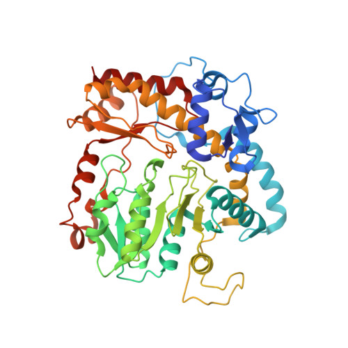
7KZD

Crystal structure of KabA from Bacillus cereus UW85 in complex with the reduced internal aldimine and with bound Glutarate


Experimental Data Snapshot

  • Method: X-RAY DIFFRACTION
  • Resolution: 1.90 Å
  • R-Value Free: 0.249 
  • R-Value Work: 0.207 
  • R-Value Observed: 0.209 

Starting Model: experimental
View more details

wwPDB Validation   3D Report Full Report


Ligand Structure Quality Assessment 


This is version 1.3 of the entry. See complete history


Literature

Snapshots along the catalytic path of KabA, a PLP-dependent aminotransferase required for kanosamine biosynthesis in Bacillus cereus UW85.

Prasertanan, T.Palmer, D.R.J.Sanders, D.A.R.

(2021) J Struct Biol 213: 107744-107744

  • DOI: https://doi.org/10.1016/j.jsb.2021.107744
  • Primary Citation of Related Structures:  
    7KZ3, 7KZ5, 7KZ6, 7KZD

  • PubMed Abstract: 

    Kanosamine is an antibiotic and antifungal monosaccharide. The kanosamine biosynthetic pathway from glucose 6-phosphate in Bacillus cereus UW85 was recently reported, and the functions of each of the three enzymes in the pathway, KabA, KabB and KabC, were demonstrated. KabA, a member of a subclass of the VI β family of PLP-dependent aminotransferases, catalyzes the second step in the pathway, generating kanosamine 6-phosphate (K6P) using l-glutamate as the amino-donor. KabA catalysis was shown to be extremely efficient, with a second-order rate constant with respect to K6P transamination of over 10 7 M -1 s -1 . Here we report the high-resolution structure of KabA in both the PLP- and PMP-bound forms. In addition, co-crystallization with K6P allowed the structure of KabA in complex with the covalent PLP-K6P adduct to be solved. Co-crystallization or soaking with glutamate or 2-oxoglutarate did not result in crystals with either substrate/product. Reduction of the PLP-KabA complex with sodium cyanoborohydride gave an inactivated enzyme, and crystals of the reduced KabA were soaked with the l-glutamate analog glutarate to mimic the KabA-PLP-l-glutamate complex. Together these four structures give a complete picture of how the active site of KabA recognizes substrates for each half-reaction. The KabA structure is discussed in the context of homologous aminotransferases.


  • Organizational Affiliation

    Department of Chemistry, University of Saskatchewan, Saskatoon, SK S7N 5C9, Canada.


Macromolecules
Find similar proteins by:  (by identity cutoff)  |  3D Structure
Entity ID: 1
MoleculeChains Sequence LengthOrganismDetailsImage
Aminotransferase class I/II-fold pyridoxal phosphate-dependent enzyme
A, B, C, D, E
A, B, C, D, E, F, G, H
445Bacillus cereusMutation(s): 0 
Gene Names: kabAGE376_30835
EC: 2.6.1.1
UniProt
Find proteins for C0JRF5 (Bacillus cereus)
Explore C0JRF5 
Go to UniProtKB:  C0JRF5
Entity Groups  
Sequence Clusters30% Identity50% Identity70% Identity90% Identity95% Identity100% Identity
UniProt GroupC0JRF5
Sequence Annotations
Expand
  • Reference Sequence
Small Molecules
Ligands 3 Unique
IDChains Name / Formula / InChI Key2D Diagram3D Interactions
PLR (Subject of Investigation/LOI)
Query on PLR

Download Ideal Coordinates CCD File 
DA [auth C]
DB [auth G]
I [auth A]
KA [auth D]
KB [auth H]
DA [auth C],
DB [auth G],
I [auth A],
KA [auth D],
KB [auth H],
Q [auth B],
QA [auth E],
YA [auth F]
(5-HYDROXY-4,6-DIMETHYLPYRIDIN-3-YL)METHYL DIHYDROGEN PHOSPHATE
C8 H12 N O5 P
RBCOYOYDYNXAFA-UHFFFAOYSA-N
GUA (Subject of Investigation/LOI)
Query on GUA

Download Ideal Coordinates CCD File 
CA [auth C],
JA [auth D],
RA [auth E],
ZA [auth F]
GLUTARIC ACID
C5 H8 O4
JFCQEDHGNNZCLN-UHFFFAOYSA-N
EDO
Query on EDO

Download Ideal Coordinates CCD File 
AA [auth B]
AB [auth F]
BA [auth B]
BB [auth F]
CB [auth F]
AA [auth B],
AB [auth F],
BA [auth B],
BB [auth F],
CB [auth F],
EA [auth C],
EB [auth G],
FA [auth C],
FB [auth G],
GA [auth C],
GB [auth G],
HA [auth C],
HB [auth G],
IA [auth C],
IB [auth G],
J [auth A],
JB [auth G],
K [auth A],
L [auth A],
LA [auth D],
LB [auth H],
M [auth A],
MA [auth D],
MB [auth H],
N [auth A],
NA [auth D],
NB [auth H],
O [auth A],
OA [auth D],
P [auth A],
PA [auth D],
R [auth B],
S [auth B],
SA [auth E],
T [auth B],
TA [auth E],
U [auth B],
UA [auth E],
V [auth B],
VA [auth E],
W [auth B],
WA [auth E],
X [auth B],
XA [auth E],
Y [auth B],
Z [auth B]
1,2-ETHANEDIOL
C2 H6 O2
LYCAIKOWRPUZTN-UHFFFAOYSA-N
Experimental Data & Validation

Experimental Data

  • Method: X-RAY DIFFRACTION
  • Resolution: 1.90 Å
  • R-Value Free: 0.249 
  • R-Value Work: 0.207 
  • R-Value Observed: 0.209 
  • Space Group: P 1
Unit Cell:
Length ( Å )Angle ( ˚ )
a = 92.94α = 108.84
b = 93.58β = 92.33
c = 106.89γ = 90.08
Software Package:
Software NamePurpose
PHENIXrefinement
PDB_EXTRACTdata extraction
MOLREPphasing
AutoProcessdata reduction
AutoProcessdata scaling

Structure Validation

View Full Validation Report


Ligand Structure Quality Assessment 


Entry History & Funding Information

Deposition Data


Funding OrganizationLocationGrant Number
Natural Sciences and Engineering Research Council (NSERC, Canada)Canada--

Revision History  (Full details and data files)

  • Version 1.0: 2021-05-26
    Type: Initial release
  • Version 1.1: 2021-06-02
    Changes: Database references
  • Version 1.2: 2023-10-18
    Changes: Data collection, Database references, Refinement description
  • Version 1.3: 2024-03-13
    Changes: Source and taxonomy