7MZM

SARS-CoV-2 receptor binding domain bound to Fab PDI 215


Experimental Data Snapshot

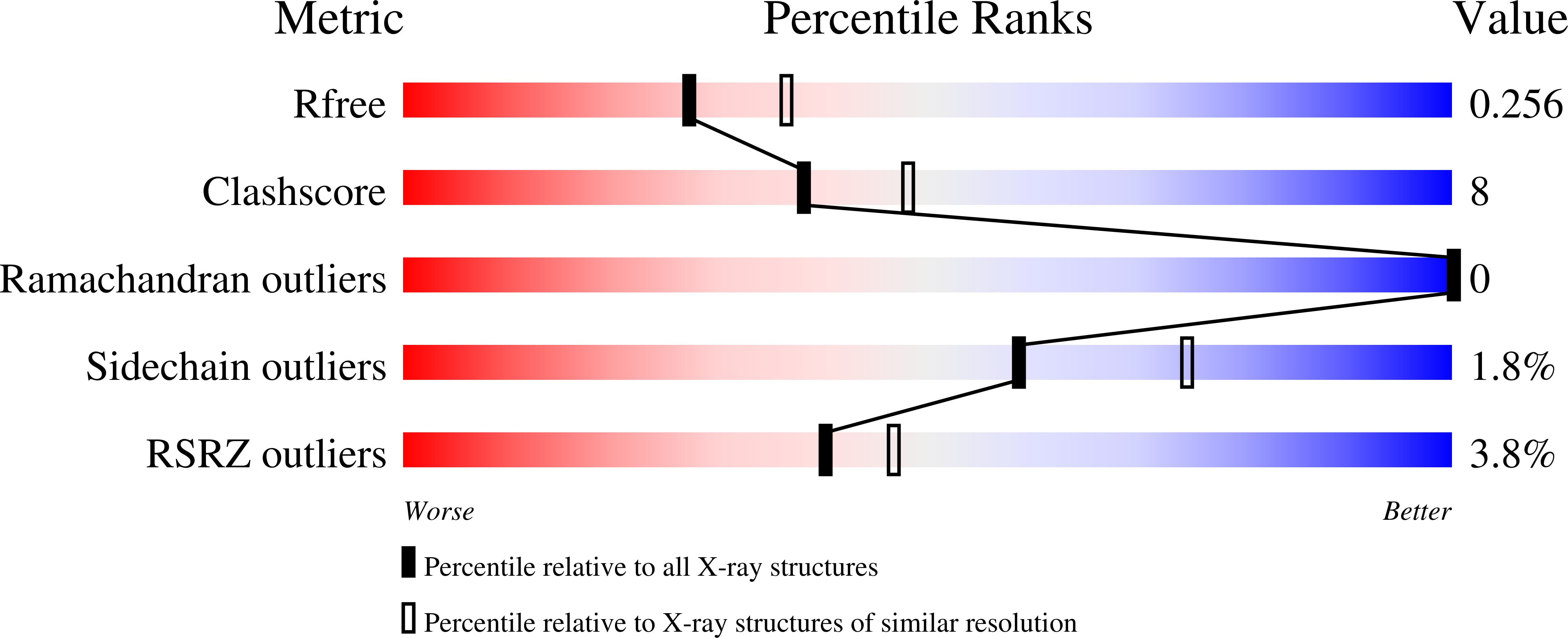
  • Method: X-RAY DIFFRACTION
  • Resolution: 2.30 Å
  • R-Value Free: 0.257 
  • R-Value Work: 0.216 
  • R-Value Observed: 0.218 

Starting Model: experimental
View more details

wwPDB Validation   3D Report Full Report


This is version 1.3 of the entry. See complete history


Literature

Landscape of human antibody recognition of the SARS-CoV-2 receptor binding domain.

Wheatley, A.K.Pymm, P.Esterbauer, R.Dietrich, M.H.Lee, W.S.Drew, D.Kelly, H.G.Chan, L.J.Mordant, F.L.Black, K.A.Adair, A.Tan, H.X.Juno, J.A.Wragg, K.M.Amarasena, T.Lopez, E.Selva, K.J.Haycroft, E.R.Cooney, J.P.Venugopal, H.Tan, L.L.O Neill, M.T.Allison, C.C.Cromer, D.Davenport, M.P.Bowen, R.A.Chung, A.W.Pellegrini, M.Liddament, M.T.Glukhova, A.Subbarao, K.Kent, S.J.Tham, W.H.

(2021) Cell Rep 37: 109822-109822

  • DOI: https://doi.org/10.1016/j.celrep.2021.109822
  • Primary Citation of Related Structures:  
    7MZF, 7MZG, 7MZH, 7MZI, 7MZJ, 7MZK, 7MZL, 7MZM, 7MZN, 7RR0

  • PubMed Abstract: 

    Potent neutralizing monoclonal antibodies are one of the few agents currently available to treat COVID-19. SARS-CoV-2 variants of concern (VOCs) that carry multiple mutations in the viral spike protein can exhibit neutralization resistance, potentially affecting the effectiveness of some antibody-based therapeutics. Here, the generation of a diverse panel of 91 human, neutralizing monoclonal antibodies provides an in-depth structural and phenotypic definition of receptor binding domain (RBD) antigenic sites on the viral spike. These RBD antibodies ameliorate SARS-CoV-2 infection in mice and hamster models in a dose-dependent manner and in proportion to in vitro, neutralizing potency. Assessing the effect of mutations in the spike protein on antibody recognition and neutralization highlights both potent single antibodies and stereotypic classes of antibodies that are unaffected by currently circulating VOCs, such as B.1.351 and P.1. These neutralizing monoclonal antibodies and others that bind analogous epitopes represent potentially useful future anti-SARS-CoV-2 therapeutics.


  • Organizational Affiliation

    Department of Microbiology and Immunology, University of Melbourne, the Peter Doherty Institute for Infection and Immunity, Melbourne, VIC 3000, Australia; Australian Research Council Centre for Excellence in Convergent Bio-Nano Science and Technology, University of Melbourne, Melbourne, VIC 3010, Australia.


Macromolecules
Find similar proteins by:  (by identity cutoff)  |  3D Structure
Entity ID: 1
MoleculeChains Sequence LengthOrganismDetailsImage
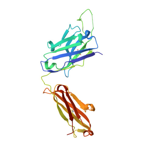
PDI 215 heavy chainA [auth H]239Homo sapiensMutation(s): 0 
Entity Groups  
Sequence Clusters30% Identity50% Identity70% Identity90% Identity95% Identity100% Identity
Sequence Annotations
Expand
  • Reference Sequence
Find similar proteins by:  (by identity cutoff)  |  3D Structure
Entity ID: 2
MoleculeChains Sequence LengthOrganismDetailsImage
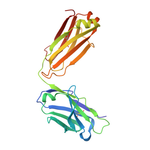
PDI 215 light chainB [auth L]216Homo sapiensMutation(s): 0 
Entity Groups  
Sequence Clusters30% Identity50% Identity70% Identity90% Identity95% Identity100% Identity
Sequence Annotations
Expand
  • Reference Sequence
Find similar proteins by:  (by identity cutoff)  |  3D Structure
Entity ID: 3
MoleculeChains Sequence LengthOrganismDetailsImage
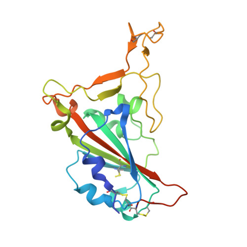
Spike protein S1C [auth A]205Severe acute respiratory syndrome coronavirus 2Mutation(s): 0 
Gene Names: S2
UniProt
Find proteins for P0DTC2 (Severe acute respiratory syndrome coronavirus 2)
Explore P0DTC2 
Go to UniProtKB:  P0DTC2
Entity Groups  
Sequence Clusters30% Identity50% Identity70% Identity90% Identity95% Identity100% Identity
UniProt GroupP0DTC2
Glycosylation
Glycosylation Sites: 1Go to GlyGen: P0DTC2-1
Sequence Annotations
Expand
  • Reference Sequence
Oligosaccharides

Help

Entity ID: 4
MoleculeChains Length2D Diagram Glycosylation3D Interactions
alpha-L-fucopyranose-(1-6)-2-acetamido-2-deoxy-beta-D-glucopyranoseD [auth B]2N-Glycosylation
Glycosylation Resources
GlyTouCan:  G86851RC
GlyCosmos:  G86851RC
GlyGen:  G86851RC
Experimental Data & Validation

Experimental Data

  • Method: X-RAY DIFFRACTION
  • Resolution: 2.30 Å
  • R-Value Free: 0.257 
  • R-Value Work: 0.216 
  • R-Value Observed: 0.218 
  • Space Group: C 2 2 21
Unit Cell:
Length ( Å )Angle ( ˚ )
a = 95.801α = 90
b = 120.325β = 90
c = 128.358γ = 90
Software Package:
Software NamePurpose
XSCALEdata scaling
PHENIXrefinement
PDB_EXTRACTdata extraction
XDSdata reduction
PHASERphasing

Structure Validation

View Full Validation Report


Entry History & Funding Information

Deposition Data


Funding OrganizationLocationGrant Number
National Health and Medical Research Council (NHMRC, Australia)AustraliaGNT2002073

Revision History  (Full details and data files)

  • Version 1.0: 2021-10-06
    Type: Initial release
  • Version 1.1: 2021-10-20
    Changes: Database references
  • Version 1.2: 2021-10-27
    Changes: Database references
  • Version 1.3: 2023-10-18
    Changes: Data collection, Refinement description